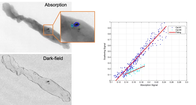
Microcalcifications consist of tiny calcium deposits, although they are usually benign, they are often present in areas of accelerated cell turnover, indicative for precancerous changes or early invasive breast cancer. Therefore, microcalcifications could act as indicators to tell whether an associated lesion is benign or malignant. The ability to non-invasively discriminate between different microcalcifications can improve the diagnostic assessment by reducing the need for biopsies. The dark-field contrast provided by grating interferometry (GI) is sensitive to the microstructures of these tiny calcium deposits and reveal insights beyond the access of current mammography technology. The team of X-ray tomography group, in collaboration with Kantonsspital Baden, has carried out a reader study to investigative whether GI can discriminate benign and malignant microcalcifications and the results show the potential of GI in non-invasively characterizing microcalcifications and aiding in the discrimination of benign from malignant lesions in clinical settings.
Original Publication
Can grating interferometry-based mammography discriminate benign from malignant microcalcifications in fresh biopsy samples?, Forte S, Wang Z, Arboleda C, Lång K, Singer G, Kubik-Huch R & Stampanoni M, European Journal of Radiology 129, 109077(2020).
Contacts
Dr. Zhentian Wang
Senior Scientist
Paul Scherrer Institut
Telephone: +41 56 310 5819
E-mail: zhentian.wang@psi.ch
