
COVID 19 research
in the BIO divison
One of our teams (Dr. R. Benoit) is specialized in the design of protein nanoparticles, using building blocks found in nature (molecular biomimetics). Virus capsids, for example, are naturally occurring protein nanoparticles that exist in a wide variety of shapes and sizes. Many copies of only few types of protein subunits spontaneously assemble to form these viral envelopes. Through recombinant DNA technology, other proteins or peptides can be fused to the subunits, resulting in the display of the attachments on the nanoparticle surface. This technology can be extremely useful for many applications. For example, engineered virus-like particles with optimized stability and epitope exposure can function as improved vaccines. Unlike viruses, the particles do not contain viral genetic material and they are therefore non-infectious. This also makes them ideal tools for the development of novel methods, for example for the specific detection of viruses.
(Foto: Mechanobiology Institute, NUS, Singapore/Melanie Lee)
Why Covid-19 hits older people particularly hard
German edition of the interview with Prof. Dr. G.V. Shivashankar (English edition to follow). For further information, please click here.
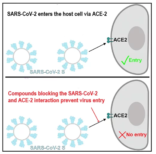
SARS-CoV-2/ACE-2 pseudovirus entry blocking assay
The spike glypcoprotein S of SARS-CoV-2 uses the human Angiotensin Converting Enzyme 2 (ACE2) as an entry receptor and recognizes it with a similar affinity to the 2002–2003 SARS-CoV isolates, which suggests it can spread efficiently in humans, in agreement with the numerous SARS-CoV-2 human-to-human transmission events reported to date (Walls et al., 2020).
For further information, please click here.
Tomographic studies of CoV-19 cell recognition
The group of Prof. Takashi Ishikawa is specialized in cryo-electron tomography and cryo X-ray tomography of cilia, small hair-like protrusions on the surface of many cell types, with important functions, such as the clearing pathogens from the lung.
For further information, please click here.