
Transmission Electron Microscopes
JEOL JEM-ARM200F
This scanning transmission electron microscope is equipped with a cold field emission gun (cold FEG), a Cs aberration corrector (ASCOR), a variety of STEM detectors (ADF, BF, ABF), multi-channel plate for BSE/SE detection, in-situ camera OneView IS (GATAN), two EDX detectors and hybrid pixel detector (JUNGFRAU) developed in-house. Single tilt and double-tilt room temperature holders are available. Single tilt cryo holders are compatible for investigations with low temperature.

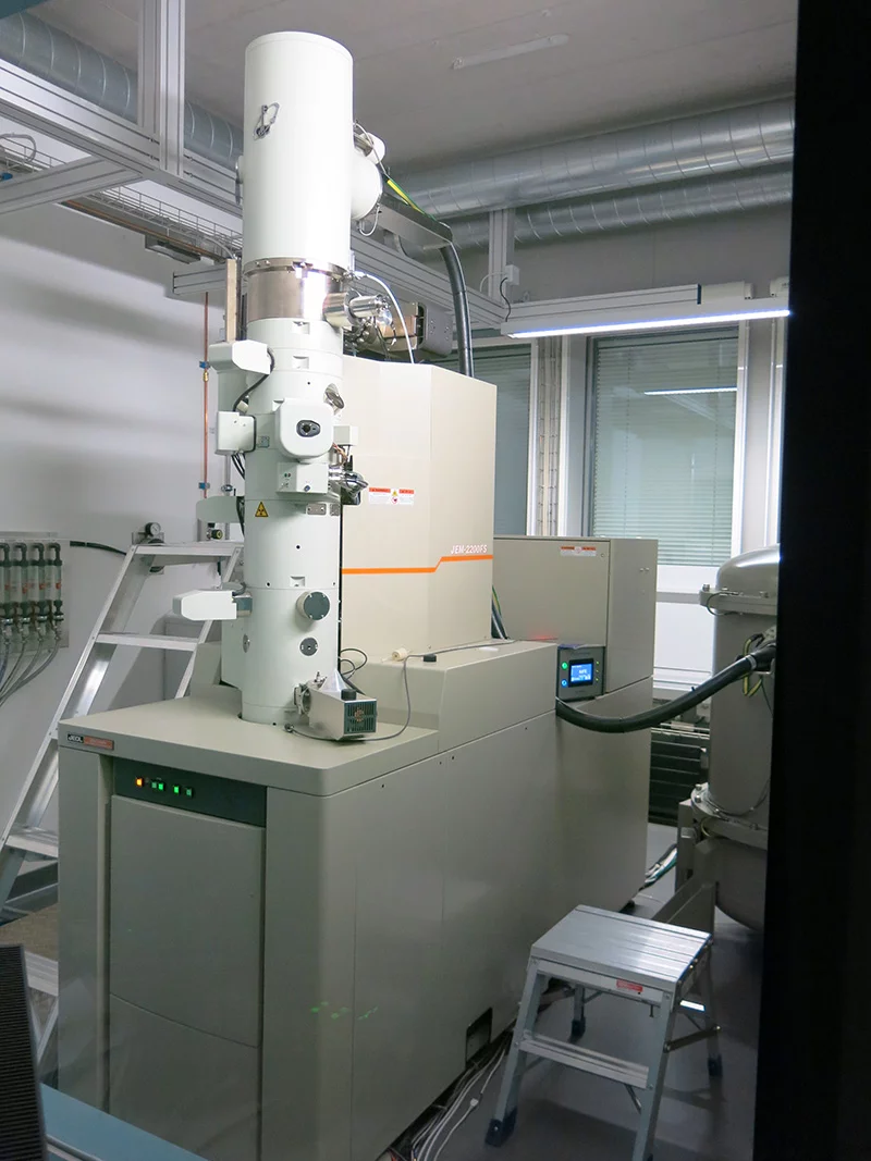
JEOL JEM 2200FS
This cryo-transmission electron microscope is equipped with a Schottky field emission gun, a tomography stage, an in-column filter and a K2 direct electron detector (GATAN). With its cryo-shields and the high-tilt pole piece it is optimized for imaging of biological samples. Single tilt room temperature and cryo holders as well as room temperature double tilt holders are available.
JEOL JEM 2010
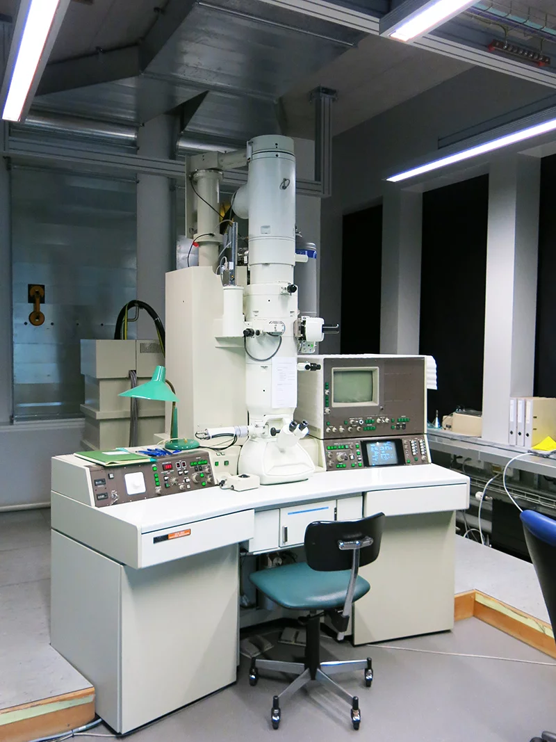
This transmission electron microscope is dedicated for materials science investigations and observation of biological samples at room temperature. It is equipped with a LaB6 cathod, a high-resolution pole piece, an EDX-detector (Oxford) and an EELS spectrometer (Gatan, Enfina). Single tilt, double tilt, cooling, heating and stretching holders are available.
FIB/SEM
ZEISS NVision40
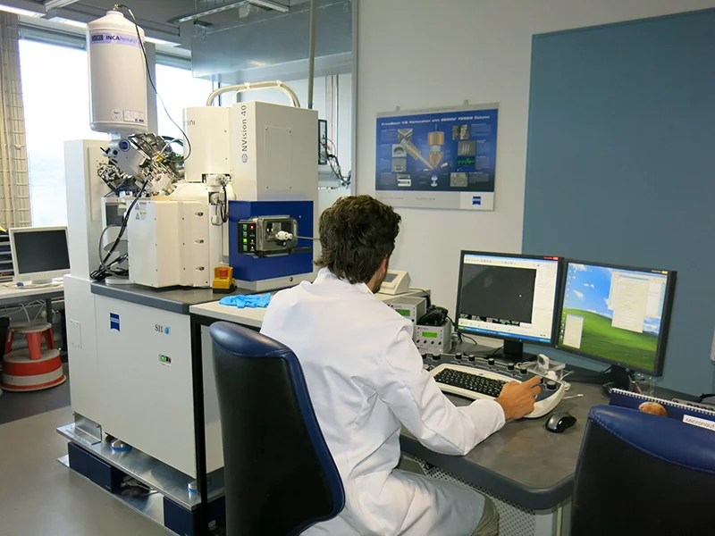
This cross-beam focused ion beam system (Schottky field emission gun for the electron column) is equipped with ESB, SE2, inlens, QBSE, STEM and EDX (Oxford) detectors, a gas injection system (SiO, Pt, C and H2O vapor), a micromanipulator (Keindiek) as well as with a cryo shuttle (VCT100, Leica) and cryo stage (LEICA) for treatment of frozen samples in the FIB and for their transfer to other instruments.
Preparation tools
Techniques available at EMF
JEM-ARM200F
- imaging including low dose and HRTEM
- selected area electron diffraction, convergent beam electron diffraction
- hollow cone illumination (conical DF)
- high resolution STEM (ADF, BF, ABF, e-ABF)
- low mag STEM
- SE/BSE
- atomic resolution EDX
- room temperature and cryo-electron tomography
- automation in TEM with SerialEM
- variety of room temperature holders as well as a compatibility with cryo holders
Requirements for samples: the samples need to be 3mm in diameter, clean, thin (some ten to some hundred nanometers, if possible close to the specimen center). The exact values depend on the technique. The observation direction is typically close to the normal axis of the sample. Magnetic samples are allowed to be inserted into a TEM only with a holder with screw fixation of the specimen.
JEM 2200FS
- imaging including low dose and HRTEM
- selected area electron diffraction, convergent beam electron diffraction
- in-column omega energy filter
- cryo-electron tomography
- single particle cryo-electron microscopy
- semi- automated and automated data acquisition with SerialEM
- variety of cryo holders
- hollow cone illumination (conical DF)
- Lorentz microscopy
Requirements for samples: the samples need to be 3mm in diameter, clean, thin (some ten to some hundred nanometers, if possible close to the specimen center). The exact values depend on the technique. The observation direction is typically close to the normal axis of the sample. Magnetic samples are allowed to be inserted into a TEM only with a holder with screw fixation of the specimen.
JEM 2010
- imaging including HRTEM
- selected area electron diffraction, convergent beam electron diffraction
Requirements for samples: the samples need to be 3mm in diameter, clean, thin (some ten to some hundred nanometers, if possible close to the specimen center). The exact values depend on the technique. The observation direction is typically close to the normal axis of the sample. Magnetic samples are allowed to be inserted into a TEM only with a holder with screw fixation of the specimen.
NVision
- SEM imaging
- STEM imaging (for very thin samples)
- EDX
- Ga-ion milling
- deposition of C, Pt and SiO
- Slice & view
- micro-manipulation
- cryo shuttle (VCT100, Leica) and cryo stage (Leica)
Requirements for samples: the samples need to fit into some standard SEM holders, their hight should not exceed few centimeters. They need to be clean. If they are non-conductive, a coating with C or some metal (typically Au, Pt or C) is required to avoid charging. If they are magnetic, they need to be very carefully fixed on the holder.