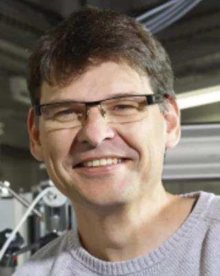
Biography
Daniel Grolimund is Senior Scientist and group leader within the Photon Science Division at the Paul Scherrer Institute. He is heading the Chemical Imaging group and leading the microXAS Beamline at the Swiss LIght Source. He received all his academic degrees from the Swiss Federal Institute of Technology Zurich (ETHZ, Switzerland), including a Diploma (dipl. natw. ETH [MSc equivalent]) and a PhD in Environmental Sciences. His PhD research focused on reactive contaminant transport in subsurface systems with special emphasis on interfacial chemistry and the role of colloidal particles as fast contaminant carriers. Following his PhD, he moved on as postdoctoral Fellow to Stanford University (Department of Geological & Environmental Sciences and Stanford Synchrotron Radiation Laboratory; Palo Alto, CA, USA). In 1999 Daniel Grolimund was awarded one of the prestigious Governor’s Lectureships of the Imperial College of Science, Technology and Medicine (London, UK). He joined the Department of Civil & Environmental Engineering as Assistant Professor (Lecturer) in Environmental Sciences and Environmental Chemistry. Already in 2000 he decided to resign to accept an unique opportunity back in Switzerland at the Swiss Light Source hosted by the Paul Scherrer Institute. Since 2006 Daniel Grolimund is scientific group leader and project responsible of the microXAS Beamline Project. Honors include the Fritz Scheffer Prize of the German Soil Science Society, a Novartis Fellowship, or the Governor’s Lectureship of the Imperial College London (UK).
Institutional Responsibilities
As project leader of the microXAS beamline at the Swiss Light Source, Daniel Grolimund is responsible for the management, coordination and operation as well as the strategic development of this hard x-ray microprobe facility. He is heading the research program of the Chemical Imaging group and is deeply involved in collaborative research projects with internal (PSI) and external academic user groups. He takes educational responsibility as supervisior of PhD students and postdoctoral fellows, including two recent PSI FELLOWs. He served frequently in synchrotron related review committee and beamline advisory panels. Between 2012 and 2015 he acted as (informal) deputy laboratory head of the Laboratory for Catalysis and Sustainable Chemistry (now LSF).
Scientific Research
The scientific research activities of Daniel Grolimund are all related to the general subject of “Imaging Chemistry in Space and Time”. His research activities are based on the application of advanced synchrotron beam and neutron beam based microscopic techniques to analyse the chemical (and physical) structure and related chemical reactivity of heterogeneous system in three dimensions – in-situ and therefore undisturbed, and, additionally, time resolved. His research activities link to the development of an advanced understanding of macroscopic reactivity and dynamics of complex systems based on the prevailing micro/nanoscopic physical and chemical characteristics revealed by high spatial resolution chemical imaging. The focus subject corresponds to enlightening our understanding of environmental chemical processes – including their link to physics, biology, and human health. Special emphasis is devoted to relate chemical micro-heterogeneities to reactive transport phenomena.
Selected Publications
For an extensive overview we kindly refer you to the Digital Object Repository at PSI (DORA) or alternatively on ORCiD and ResearcherID. Publications associated with the microXAS beamline can be found here.
Some recent publication highlights include:
Nickel Poisoning of a Cracking Catalyst Unravelled by Single Particle X-ray Fluorescence-Diffraction-Absorption Tomography, M. Gambino, M. Veselý, Ma. Filez, R. Oord, D. Ferreira Sanchez, D. Grolimund, N. Nesterenko, D. Minoux, M. Maquet, F. Meirer, B.M. Weckhuysen, Angewandte Chemie, 59/10, 3922-3927, (2020).
Operando X-ray diffraction during laser 3D printing, S. Hocine, H. Van Swygenhoven, S. Van Petegem, C.S.T. Chang, T. Maimaitiyili, G. Tinti, D.F. Sanchez, D. Grolimund, N. Casati, Materials Today, 34, 30-40, (2020).
Towards time-resolved laser T-jump/X-ray probe spectroscopy in aqueous solutions, O. Cannelli, C. Bacellar, R.A. Ingle, R. Bohinc, D. Kinschel, B. Bauer, D. S. Ferreira, D. Grolimund, G.F. Mancini, M. Chergui, Structural Dynamics, 6/6, 064303, (2019).
Spatial displacement of forward diffracted X-ray beam by perfect crystals, A. Rodriguez-Fernandez, V. Esposito, D.F. Sanchez, K.D. Finkelstein, P. Jurianic, U. Staub, D. Grolimund, S. Reiche, B. Pedrini, Acta Crystallographica Section A, 74/2, 75-87, (2018).
Fast Novel 2D/3D Microanalysis by Energy Dispersive X-ray Absorption Spectroscopy Tomography, D. Ferreira Sanchez, A.S. Simionovici, L. Lemelle, V. Cuartero Yague, O. Mathon, S. Pascarelli, A. Bonnin, R. Shapiro, K. Konhauser, D. Grolimund, P. Bleuet, Sci. Rep., 7, 16453, (2017).
Unraveling heme detoxification and crystallization in the malaria parasite by correlative X-ray microscopy, S. Kapishnikov, D. Grolimund, G. Schneider, E. Pereiro, J. McNally, J. Als-Nielsen, L. Leiserowitz, Sci. Rep., 7, 7610, (2017).
Geochemical Interactions of Plutonium(VI) with Opalinus Clay Studied by Spatially Resolved Synchrotron Radiation Techniques, U. Kaplan, S. Amayri, D.R. Fröhlich, J. Drebert, D. Grolimund, C. Borca, T. Reich, Environ. Sci. Technol., 51, 7892-7902, (2017).
Microscopic Chemical Imaging: A Key to Understand Ion Mobility in Tight Formations, D. Grolimund, H.A.O Wang, L.R. Van Loon, F. Marone, N. Diaz, A. Kaestner, A. Jakob, CMS Workshop Lecture Series, Chapter 9, 105-128, in: Filling the Gaps – from Microscopic Pore Structures to Transport Properties in Shales (T. Schäfer, R. Dohrmann, and H.C. Greenwell, editors). CMS Workshop Lectures, 21. The Clay Minerals Society, Chantilly, Virginia, USA. ISBN - 978-1-881208-46-4, (2016).
Mother plant-mediated pumping of zinc into the developing seed, L.I. Olsen, T.H. Hansen, C. Larue, J.T. Østerberg, R.D. Hoffmann, J. Liesche, U. Krämer, S. Surble, S. Cadarsi, V.A. Samson, D. Grolimund, S. Husted, M. Palmgren, NATURE Plants, 16036, (2016).
Personal cornerstone publications include:
Mimicking additive manufacturing conditions combining laser melting of Ti-6Al-4V with in situ high-speed X-ray diffraction, C. Kenel, D. Grolimund, X. Li, E. Panepucci, V.A. Samson, D. Ferreira Sanchez, F. Marone, C. Leinenbach, Sci. Rep., 7, 16358, (2017).
Rapid Mapping of Lithiation Dynamics in Transition Metal Oxides using Operando X-Ray Absorption Spectroscopy, L. Nowack, D. Grolimund, V. Samson, F. Marone, V. Wood, Sci. Rep., 6, 21479, (2016).
Barite precipitation following celestite dissolution in reactive transport experiments: a SEM/BSE and µ-XRD/XRF study, J. Poonoosamy, E. Curti, G. Kosakowski, L. R. Van Loon, D. Grolimund & U. Mäder, Geochim. Cosmochim. Acta, 182, 131-144, (2016).
Application of micro X-ray diffraction to investigate the reaction products formed by the alkali-silica reaction in concrete structures, R. Dähn, A. Arakcheeva, Ph. Schaub, P. Pattison, G. Chapuis, D. Grolimund, E. Wieland, A. Leemann, Cement and Concrete Research, 79, 49-56, (2016).
Characterization of 79Se in UO2 spent nuclear fuel by micro X-ray absorption spectroscopy and its thermodynamic stability, E. Curti, A. Puranen, D. Grolimund, D. Jädernas, D. Cheptiakov, A. Mesbah, J.A. Ibers, Environmental Science: Processes & Impacts, 17 (10), 1760-1768, (2015).
Kinoform diffractive lenses for efficient nano-focusing of hard X-rays, P. Karvinen, D. Grolimund, M. Willimann, B. Meyer, M. Birri, C. Borca, J. Patommel, G. Wellenreuther, G. Falkenberg, M. Guizar-Sicairos, A. Menzel, C. David, Optics Express, 22/14, 16676-16685, (2014).
Simultaneous visualization of 32 proteins and protein modifications in formalin-fixed paraffin-embedded tissues with subcellular resolution by imaging mass cytometry, C. Giesen, H.A.O Wang, P. Schüffler, B. Hattendorf, D. Grolimund, A. Jacobs, N. Zivanovic, J.M. Buhmann, S. Brandt, Z. Varga, P.J. Wild, D. Günther, B. Bodenmiller, Nature Methods, 11/4, 417-423, (2014).
Quantitative Chemical Imaging of Element Diffusion into Heterogeneous Media Using Laser Ablation Inductively Coupled Plasma Mass Spectrometry, Synchrotron Micro-X-ray Fluorescence, and Extended X-ray Absorption Fine Structure Spectroscopy, H.A.O. Wang, D. Grolimund, D. Guenther, L. von Loon, K. Barmettler, C.N. Borca, B. Aeschlimann, Analytical Chemistry, 16, 6259–6266, (2011).
Picosecond Time-Resolved X-Ray Emission Spectroscopy: Ultrafast Spin-State Determination in an Iron Complex, G. Vanko, P. Glatzel, V-T. Pham, R. Abela, D. Grolimund, C.N. Borca, S.L. Johnson, C.J. Milne, Ch. Bressler, Angew. Chemie, 49(34), 5910-5912, (2010).
Femtosecond XANES of the light-induced spin cross-over in a Iron(II) complex, Ch. Bressler, C. Milne, V.-T. Pham, A. ElNahhas, R.M. van der Veen, W. Gawelda, S. Johnson, P. Beaud, D. Grolimund, M. Kaiser, C.N. Borca, G. Ingold, R. Abela, M. Chergui, SCIENCE, 323, 489–492, (2009).
Time resolved Laue diffraction of deforming micropillars, R. Maaß, S. Van Petegem, H. Van Swygenhoven, P.M. Derlet, C.A. Volkert, D. Grolimund, Phys. Rev. Lett., 99 (14): Art. No. 145505, (2007).
Spatiotemporal Stability of Femtosecond Hard X-ray Undulator Radiation Studied by Control of Coherent Optical Phonons, P. Beaud, S. L. Johnson, A. Streun, R. Abela, D. Abramsohn, D. Grolimund, F. Krasniqi, T. Schmidt, V. Schlott, G. Ingold, Phys. Rev. Lett., 99, 174801(1-4), (2007).
Identification of Cr Species at the Solution-Hematite Interface after Cr(VI)-Cr(III) Reduction using GI-XAFS and Cr L-Edge NEXAFS, D. Grolimund, T.P. Trainor, J.P. Fitts, T. Kendelewicz, P. Liu, S.A. Chambers, G.E. Brown, Jr., J. Synch. Rad., 6, 612, (1999).
Colloid Facilitated Transport of Strongly Sorbing Contaminants in Natural Porous Media: A Laboratory Column Study, D. Grolimund, M. Borkovec, K. Barmettler, H. Sticher, Research Communication, Environ. Sci. Technol., 30, 3118, (1996).
Patents
D. Guenther, D. Grolimund, H.A.O. Wang (inventors), Ablation cell for laser ablation of sample material has sample chamber which receives sample in state which enables laser beam to enter sample chamber through lateral window and lateral opening, and to impinge on sample surface, Patent Number(s): WO2014146724-A1; US2014287953-A1; AU2013382989-A1; CA2907483-A1; WO2014147260-A1; CA2907573-A1, Patent Assignee Name(s) and Code(s): ETH ZUERICH (ETHZ-Non-standard), SCHERRER INST PAUL (SCHE-Non-standard), GUENTHER D(GUEN-Individual), GROLIMUND D(GROL-Individual), WANG H(WANG-Individual). Derwent Primary Accession Number: 2014-R61808 [82], (2014).

