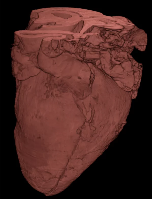
Heart failure, as well as sudden cardiac death associated with arrhythmias, are the main causes of mortality, and they are mostly associated with alteration in the muscle tissue (e.g. the presence of important fibrosis or scar). This is why the microscopic organization, as well as its integration in macro anatomy, needs to be completely understood to get a better understanding and develop adequate treatment strategies. Synchrotron X-ray Phase Contrast micro-tomography, as implemented at the TOMCAT beamline (PSI), provide essential and unique 3D reconstructions of complete hearts (see figure) for understanding cardiac microstructure and its influencing factors, both on small animals or human fetal hearts. The aim of our current Heart Imaging Project is to quantify cardiac structures and tissues for the first time at micrometre scale, non-destructively, without the use of contrast agents and in 3D. Insights on the cardiac organ morphology like the myofibres, vessels and trabeculation organization, and how they change with disease, are crucial for future clinical applications, not only to understand heart remodelling and the different patterns in cardiovascular disease, but also to provide more personalized and adapted treatments. In this project, we are working in collaboration with the University Pompeu Fabra (UPF Barcelona), the Hospital Clinic and IDIBAPS (Barcelona), the Institute Cardiovascular Sciences (UCL London) and University Hospital Centre Zagreb.
Publications
- Gonzalez-Tendero A, Zhang C, Balicevic V, Cárdenes R, Loncaric S, Butakoff C, Paun B, Bonnin A, Garcia-Cañadilla P, Muñoz-Moreno E, Gratacós E, Crispi F, Bijnens B. “Whole heart detailed and quantitative anatomy, myofibre structure and vasculature from X-ray phase-contrast synchrotron radiation-based micro-CT”. EHJ Cardiovasc Im, 2016.
- Gonzalez Tendero A., PhD Thesis, 2014.
- Balicevic, V. et al. (2015), Functional Imaging and Modeling of the Heart, pp. 111–119.
- Dejea H, Bonnin A, Garcia-Canadilla P, Stampanoni M, Bijnens B. “X-Ray Phase Contrast Imaging for Cardiac Microstructure Visualization.” École de Microscopie Fonctionnelle en Biologie, Seignosse, Sep 30 – Oct 7, 2016.
- Dejea, H., Garcia-Canadilla, P., Cook, A.C. et al. Comprehensive Analysis of Animal Models of Cardiovascular Disease using Multiscale X-Ray Phase Contrast Tomography. Sci Rep 9, 6996 (2019).
Collaboration
- Prof. Dr. Bart Bijnens, Universitat Pompeu Fabra (UPF), Barcelona, Spain
- Dr. Andrew Cook, University College London (UCL), London, United Kingdom
- Dr. Maja Cikes, Dr Ivo Planinc University Hospital Centre Zagreb, Zagreb, Croatia
- Dr. Eduard Guasch and Dr. Fatima Crispi, Hospital Clínic – IDIBAPS, Barcelona, Spain
Contacts
Dr. Patricia Garcia-Canadilla, patricia.garciac@upf.edu
Hèctor Dejea i Velardo, hector.dejea@psi.ch, +41 56 310 51 95