By combining two advanced synchrotron-based microscope techniques, an international team of scientists led by the Niels Bohr Institute (University of Copenhagen, Denmark) has managed to obtain new information about the ravaging mode of operation of malaria parasites after invading the red blood cells of humans.
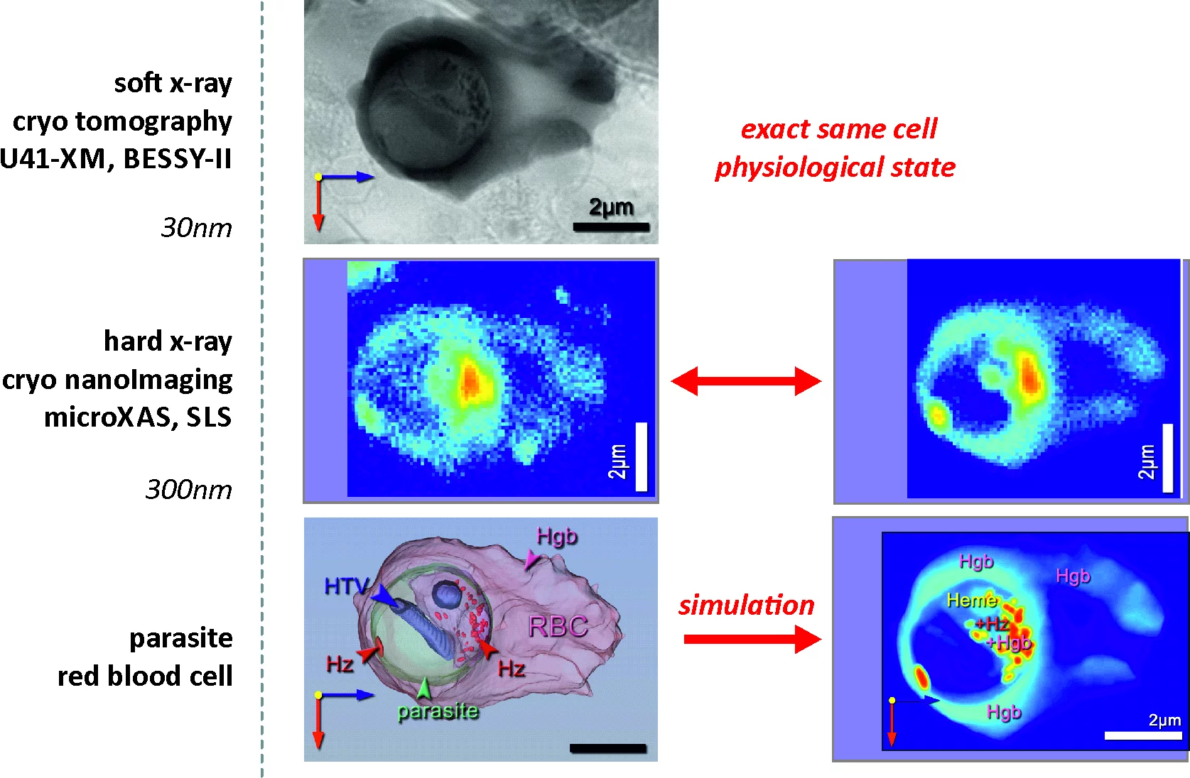
Based on a new chemical imaging methodology – in situ correlative X-ray fluorescence microscopy and soft X-ray tomography – the researchers were able to create detailed 3D images of malaria parasites in human blood cells. Structural and (bio-)chemical details are observed which have not been reported before. For the first time snapshots of in vivo states of parasites at different stages of the parasite development are recorded. Based on the discerned temporal evolution of the deterioration of the red blood cell and the related growth of the parasite, fundamental information about the digestion rate and well as the rates of related biochemical processes are obtained. This unique information reveals a strong coupling of several key processes vital to the parasites growth.
This information can be utilized when designing new medication to more effectively fight malaria - a disease claiming over 400.000 lives each year, a majority of whom are infants.
In addition to the microXAS beamline of the Swiss Light Source at Paul Scherrer Institut, the study involved the cooperation between the following international research centers: the Niels Bohr Institute from University of Copenhagen, Helmholtz Research Center from Berlin, Weizmann Institute of Science from Israel, BESSY-II, and ALBA Synchrotron.
Read the full story
Unraveling heme detoxification and crystallization in the malaria parasite by correlative X-ray microscopyS. Kapishnikov, D. Grolimund, G. Schneider, E. Pereiro, J. McNally, J. Als-Nielsen, and L. Leiserowitz
Nature Scientific Reports, 7, Article number: 7610, published online: 08 August 2017
DOI: 10.1038/s41598-017-06650-w
Contact
Dr Daniel GrolimundLaboratory for Synchrotron Radiation and Femtochemistry
Swiss Light Source, Paul Scherrer Intitute
5232 Villigen-PSI, Switzerland
Dr Sergey Kapishnikov
Xray and Neutron Science
Niels Bohr Institute, University of Copenhagen
2100 Copenhagen, Denmark