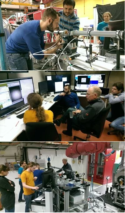
X-ray Free Electron Lasers (XFELs) combine the properties of synchrotron radiation (short wavelengths) and laser radiation (high lateral coherence, ultrashort pulse durations). These outstanding machines allow to study ultra-fast phenomena at an atomic level with unprecedented temporal resolution for answering the most intriguing open questions in biology, chemistry and physics. SwissFEL at the Paul Scherrer Institute is a large scale research XFEL facility currently under commissioning. It is designed to provide femtosecond hard x-ray pulses (2-12 keV, Aramis) at 100Hz repetition rate and in the near future also in the soft x-ray range (250eV – 2keV, Athos). Recently, a collaboration of scientists from PSI and XFEL (at DESY, Hamburg), measured for the first time the wave-front of the x-ray pulses generated at SwissFEL. This was accomplished in a single-shift beamtime using an in-house developed prototype instrument (Wave-Front Sensor) at the Bernina experimental station, a beamine specifically designed for ultra-fast pump-probe studies in condensed matter and material science.
For performing advanced ultra-fast experiments, full control of the FEL photon beam is required: from timing and synchronization (up to few femtoseconds) to spatial pointing, beam dimensions and transverse distribution. At the Bernina instrument the x-ray beam is manipulated by bendable Kirkpatrik-Baez (KB) x-ray mirrors, which can focus the photon beam to lateral dimensions as small as few micrometers. An essential aspect for successful experiments is the determination of the photon beam quality at the sample location. In fact, changes in spot dimension, shape and/or pointing may drastically alter the experimental condition in a detrimental way. For these reasons, an instrument fully dedicated to this purpose is foreseen at Bernina experimental station. Based on the Talbot effect, the single grating Wave-Front Sensor (WFS) can be used to measure the x-rays wave-front which contains important information about the focused x-rays spot size, location and shape, in just one shot.
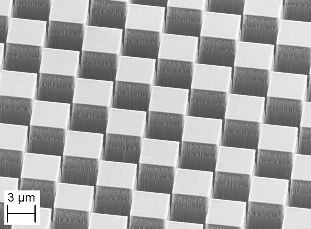
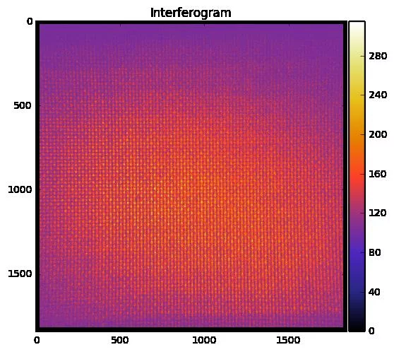
In a nutshell the WFS is composed by a phase diffraction grating placed one meter downstream of the sample location diffracting the FEL photon beam to a detector consisting of a fluorescent screen and a microscope, called x-ray eye. The x-ray eye is placed at a known distance from the grating, and collects the interferogram which is, in principle, a self image of the grating. In reality, the interferogram reflects the distortions due to the FEL wave-front shape. By measuring the displacement of the diffracted fringes it is possible to reconstruct the FEL wave-front and estimate the beam quality. In addition, the actual position of the focus as well as the beam size can be derived for each single FEL shot.
In a joint effort scientist from the SwissFEL Bernina (C. Svetina, G. Mancini, R. Mankowsky, B. Pedrini, S. Zerdane, P. Beaud, H. T. Lemke), Laboratory for Micro and Nanotechnology (F. Koch, V. Yurgens, C. David), Optics group (J. Krempasky, U. Wagner, U. Flechsig) and external collaborators from X-FEL in Hamburg (M. Ladislav, P. Vagovic, A. Mancuso) succeeded in measuring for the very first time the curvature and shape of the SwissFEL wave-front at Bernina experimental station. They have measured how the focal spot moves and how the beam size changes when manipulating the x-ray beam with the KB active focusing mirrors. Furthermore, the high resolution of the wave-front sensor was demonstrated by measuring the degradation of the wave-front introduced by controlled aberrations on the photon beam.
First results from preliminary data analysis show that the WFS can indeed be used to derive the required information for optimizing the experimental conditions. In the near future the actual instrument will be installed and commissioned. The final instrument will host a set of diffraction gratings to cover the whole Aramis SwissFEL energy range and will allow to both optimize the focus at the end-station and give support to machine studies, thus providing useful shot-to-shot information about the beam quality. The current prototype will further be optimized for ultra-fast phase contrast x-ray imaging studies. This technique can be used to probe light-induced emergent states in materials on ultrafast timescales by measuring the phase changes of x-ray pulses in transmission.
Contact
Dr. Christian SvetinaSwissFEL Bernina Group, WBBA/015
Paul Scherrer Institute
Telephone: +41 56 310 43 56
E-mail: cristian.svetina@psi.ch


