Bismuth Ferrite (BiFeO3 - BFO) is the only natural multiferroic material where the magneto-electric coupling persists at room temperature, which makes it the prototypical multiferroic material. A ferroelectric order coexists with antiferromagnetic ordering of the Fe magnetic moments. Magneto-electric coupling links the two ferroic orders and makes it is possible to alter the magnetic order by means of an electrical field. This of particular interest for technological applications, as this would avoid the use of electrical currents to address the magnetic state. In addition, the magnetic sublattices do not simply align parallel to each other in neighboring unit cells. Instead the magnetic moments are twisted in a spin cycloidal state with a period of approximately 64 nm, providing an additional point of interest for the study of this complex material.
The combined nanoscale imaging of the magnetic and ferroelectric order is a crucial requirement for the investigation of multiferroic materials. To achieve this, a combination of elemental, magnetic and ferroelectric sensitivities), along with the capability for high spatial resolution are required. The only technique able to combine all these requirements is soft X-ray ptychographic imaging. However, in order to utilize soft X-ray ptychography imaging is that the sample needs to be thin enough to allow for the transmission of soft X-ray radiation through it which is strongly absorbed by matter. This is a critical challenge for epitaxial materials such as BFO, as they need to be grown on the surface of a bulk, single-crystalline substrate.
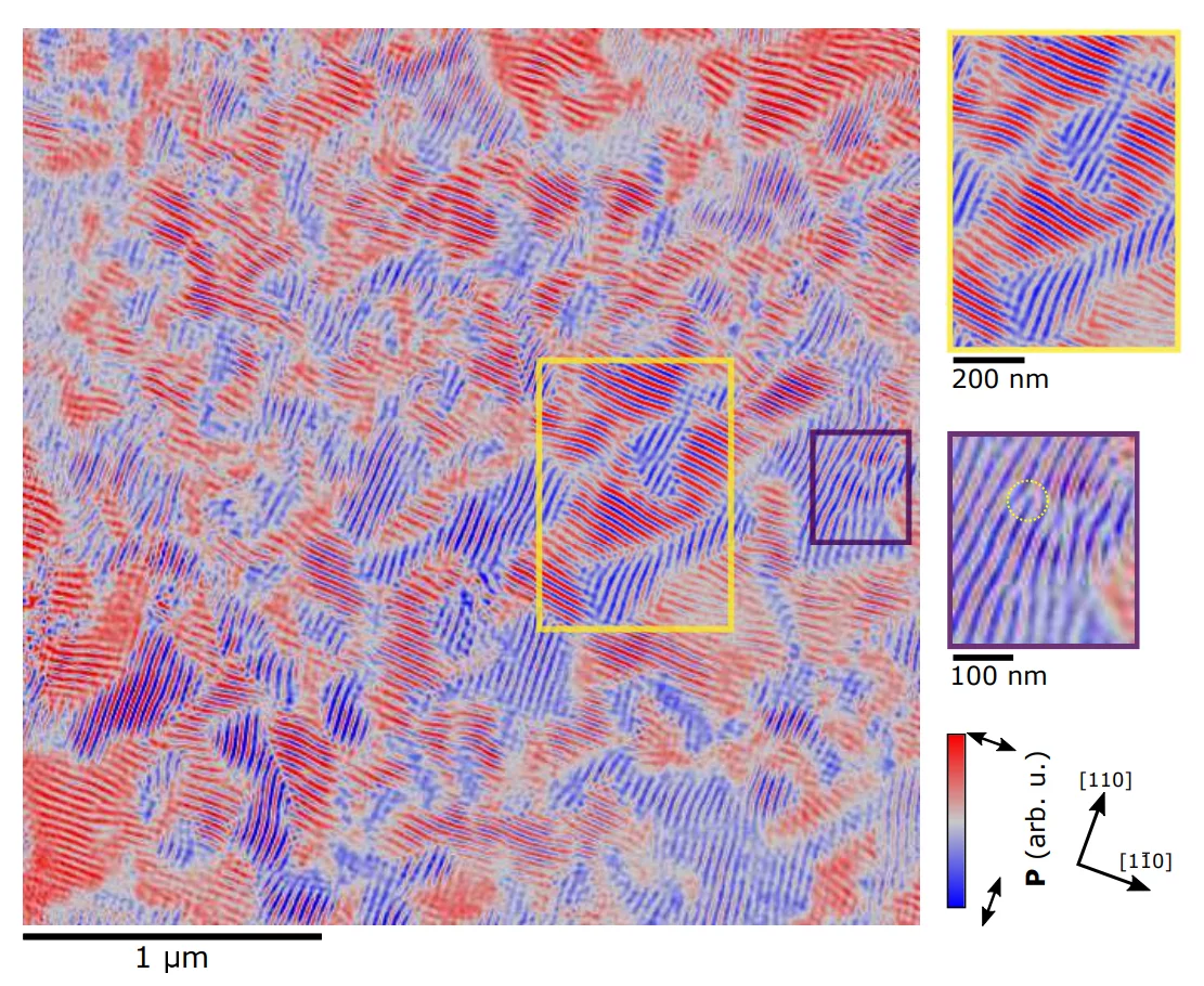
In this work, a collaboration of scientists from the Swiss Light Source and the National Cheng Kung University in Taiwan, performed soft X-ray ptychographic imaging of multiferroic domain structures in thin freestanding BFO films. These were fabricated by growing a water-soluble sacrificial layer between the BFO and the single-crystalline bulk SrTiO3 substrate. By dissolving the sacrificial layer, the BFO films could then be transferred onto any substrate of choice, which in this case was a TEM grid. The domain configuration was then visualised by soft X-ray ptychography at the L3 absorption edge of Fe, which revealed the spin cycloid as stripes in different directions on the ferroelectric domains and the perfect coupling between them.
This work, now published in the journal Advanced Materials, is the first stepping stone towards the investigation of the nanoscale dynamics in multiferroic materials, and future work will include the study of the three-dimensional structure of the ferroelectric and antiferromagnetic domains, so far only possible through destructive methods.
Contact:
Dr. Tim Alexander Butcher
Swiss Light Source
Paul Scherrer Institut
Telephone: +41 56 310 5696
E-mail: tim.butcher@psi.ch
Dr. Jörg Raabe
Swiss Light Source
Paul Scherrer Institut
Telephone: +41 56 310 5193
E-mail: joerg.raabe@psi.ch
Original Publication:
Ptychographic Nanoscale Imaging of the Magnetoelectric Coupling in Freestanding BiFeO3
Tim A. Butcher, Nicholas W. Phillips, Chun-Chien Chiu, Chia-Chun Wei, Sheng-Zhu Ho, Yi-Chun Chen, Erik Fröjdh, Filippo Baruffaldi, Maria Carulla, Jiaguo Zhang, Anna Bergamaschi, Carlos A. F. Vaz, Armin Kleibert, Simone Finizio, Jan-Chi Yang, Shih-Wen Huang, and Jörg Raabe
Advanced Materials (2024), DOI:10.1002/adma.202311157