

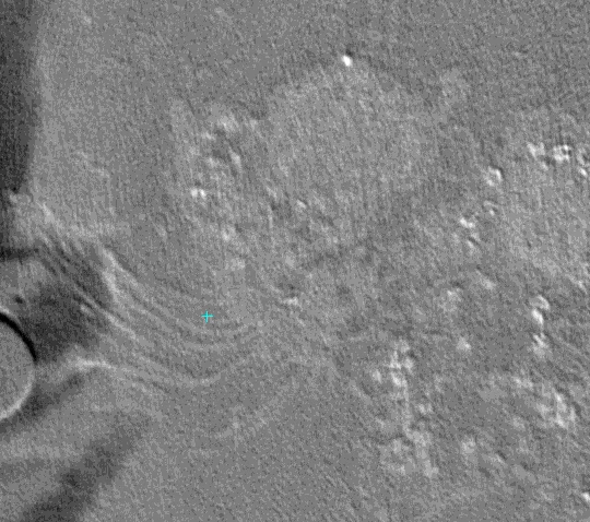
Human ciliated bronchial epithelial cell imaged by cryo X-ray tomography method (unpublished data). Ciliated bronchial epithelial cells are typical host cells which coronavirus infects. We are working on the mechanism of coronavirus targeting these cells by bio-imaging methods. The cells are cultured and prepared by the Ishikawa group at the BIO department and imaged using ptychographic tomography at the cSAXS beamline.
The group of Prof. Takashi Ishikawa is specialized in cryo-electron tomography and cryo X-ray tomography of cilia, small hair-like protrusions on the surface of many cell types, with important functions, such as the clearing pathogens from the lung. Ciliated bronchial epithelial cells are typical host cells which coronaviruses infect. We are experienced in the in vitro culture of this cell type, which allows us to produce enough cells for detailed structural experiments. Our aim is to study the mechanism that coronaviruses use for recognizing and targeting human ciliated cells, in the context of the cell, using newest generation bio-imaging methods at PSI beamlines.