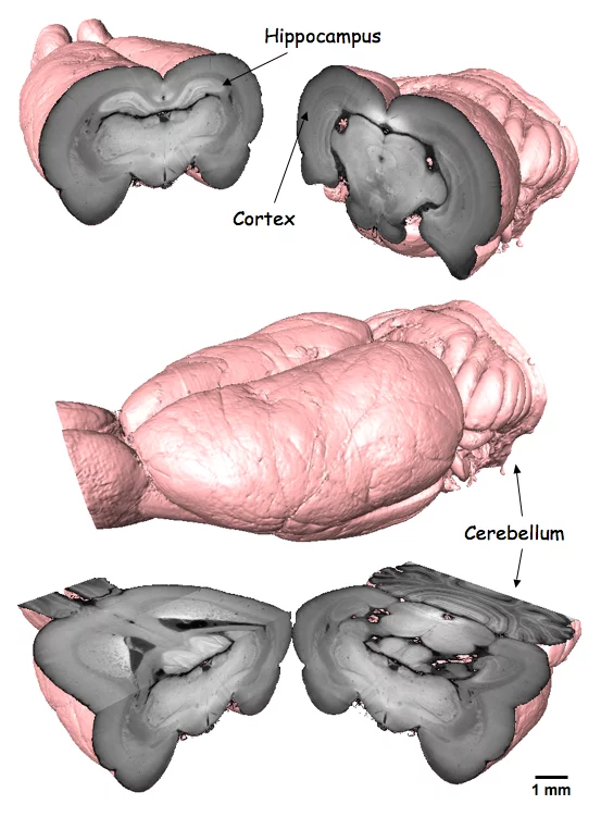
Conventional absorption based X-ray microtomography can become limited for objects showing only very weak attenuation contrast at high energies. However, a wide range of samples studied in biology and materials science can produce significant phase shifts of the X-ray beam and thus phase contrast X-ray imaging can provide substantially increased contrast sensitivity. A Differential Phase Contrast (DPC) imaging facility, based on grating interferometry, has been installed at the TOMCAT beamline, with the aim of having a high-throughput of samples in terms of fast data acquisition and post-processing. We have made hardware and software advancements to enable a range of PEC tomographic imaging methods to be applied, such as local and 'widefield' PEC tomography. Darkfield imaging, based on the mechanism of small-angle scattering, provides simultaneous and complementary information about a sample at the micron and the sub-micron length scales. The technique allows the visualisation of the soft tissue features of a rat brain, for example, with a contrast impossible to obtain with conventional absorption-based imaging.
Read full article
Read full article
Facility: SLS
Swiss Light Source, Paul Scherrer Institut, 5232 Villigen PSI, Switzerland
Email: marco.stampanoni@psi.ch
Reference
S. A. McDonald, F. Marone, C. Hintermüller, G. Mikuljan, C. David, F. Pfeiffer and M. Stampanoni, J. Synchrotron Rad. 16, 562-572 (2009).Contact
Marco StampanoniSwiss Light Source, Paul Scherrer Institut, 5232 Villigen PSI, Switzerland
Email: marco.stampanoni@psi.ch