
The beamline for TOmographic Microscopy and Coherent rAdiology experimentTs (TOMCAT) [1] is operated by the X-ray Tomography Group and offers cutting-edge technology and scientific expertise for exploiting the distinctive peculiarities of synchrotron radiation for fast, non-destructive, high resolution, quantitative investigations on a large variety of samples. Absorption-based and phase contrast imaging are routinely performed with isotropic voxel sizes ranging from 0.16 to 11 μm (fields-of-view (h x v) of 0.4 x 0.3 mm2 and 22 x 3-7 mm2, respectively) in an energy range of 8-45 keV. Phase contrast is obtained with simple edge-enhancement, propagation-based techniques [2, 3] or through grating interferometry [4].
Typical acquisition times are on the order of seconds to a few minutes. However, dynamic processes can be followed in 4D (3D space + time) using the ultra-fast endstation, which provides sub-second temporal resolution [5] for extended time periods thanks to the in-house developed GigaFRoST system [6]. A laser-based heating system [7] and a cryojet and cryo-chamber are available as standard installations and are compatible with both the standard and ultra-fast endstations. It is also possible to bring specialized, user-defined instrumentation to TOMCAT. Please contact beamline staff in advance to discuss this option.
A temporal resolution of a few (< 5) minutes can also be achieved with the hard X-ray full-field microscope setup [8] delivering a pixel size of 65 nm for microscopic samples (~75x75 μm2 field-of-view).
3D tomographic datasets are reconstructed from 2D projections using highly optimized software [9, 10] based on Fourier methods and a user-friendly interface (i.e., an ImageJ plug-in). Remote access to a flexible HPC facility is available for subsequent advanced post-processing and data quantification. A suite of analytical and iterative reconstruction routines is provided, additional ad-hoc tools can be easily installed by the single user.
Technical Data
| Energy range | 8-45 keV |
| Highest 3D spatial resolution | ca. 1 μm in parallel beam geometry ca. 200 nm in full-field geometry |
| Max. temporal resolution | 20 Hz |
| Available techniques | - Absorption-based tomographic microscopy - Propagation-based phase contrast tomographic microscopy - Ultra-fast tomographic microscopy - Grating interferometry - Absorption and phase contrast nanotomography |
| Available devices for in situ sample conditioning | - Laser-based heating system - Cryojet and cryo-chamber |
Beamline location
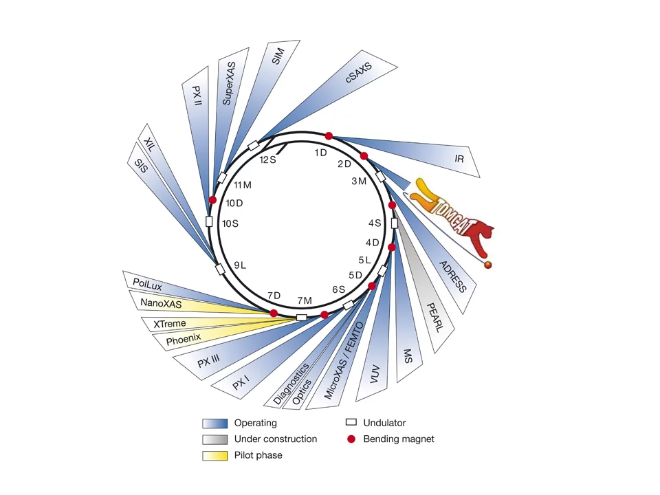
The TOMCAT beamline is located at the X02DA port of the SLS, between the Infrared and the ADRESS beamline.
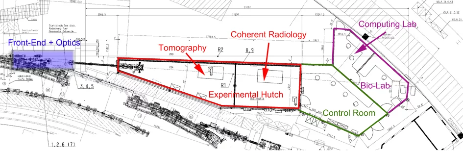
Beamline layout
References
- M. Stampanoni, A. Groso, A. Isenegger, G. Mikuljan, Q. Chen, A. Bertrand, S. Henein, R. Betemps, U. Frommherz, P. Bohler, D. Meister, M. Lange, and R. Abela, "Trends in synchrotron-based tomographic imaging: the SLS experience", Developments in X-Ray Tomography V, Proceedings of the Society of Photo-Optical Instrumentation Engineers (Spie), 6318, U199-U212 (2006). DOI: 10.1117/12.679497
- A. Groso, R. Abela, and M. Stampanoni, "Implementation of a fast method for high resolution phase contrast tomography", Optics Express, 14, 8103-8110 (2006). DOI: 10.1364/OE.14.008103
- D. Paganin, S. C. Mayo, T. E. Gureyev, P. R. Miller, and S. W. Wilkins, "Simultaneous phase and amplitude extraction from a single defocused image of a homogeneous object", Journal of Microscopy, 206, 33-40 (2002). DOI: 10.1046/j.1365-2818.2002.01010.x
- S. A. McDonald, F. Marone, C. Hintermüller, G. Mikuljan, C. David, F. Pfeiffer, and M. Stampanoni, "Advanced phase-contrast imaging using a grating interferometer", J. Synchrotron Rad., 16, 562-572 (2009). DOI: 10.1107/S0909049509017920
- R. Mokso, F. Marone, D. Haberthur, J. C. Schittny, G. Mikuljan, A. Isenegger, and M. Stampanoni, "Following Dynamic Processes by X-ray Tomographic Microscopy with Sub-second Temporal Resolution", 10th International Conference on X-Ray Microscopy, 1365, 38-41 (2011). DOI: 10.1063/1.3625299
- R. Mokso, C. M. Schlepütz, G. Theidel, H. Billich, E. Schmid, T. Celcer, et al., "GigaFRoST: The Gigabit Fast Readout System for Tomography", J. Synchrotron Rad., 24 (6), 1250-1259 (2017). DOI: 10.1107/S1600577517013522
- J. L. Fife, M. Rappaz, M. Pistone, T. Celcer, G. Mikuljan, and M. Stampanoni, "Development of a laser-based heating system for in-situ synchrotron-based x-ray tomographic microscopy", J. Synchrotron Rad., 19, 352 (2012). DOI: 10.1107/S0909049512003287
- M. Stampanoni, R. Mokso, F. Marone, J. Vila-Comamala, S. Gorelick, P. Trtik, et al., "Phase-contrast tomography at the nanoscale using hard x-rays", Physical Review B, 81, 140105R (2010). DOI: 10.1103/PhysRevB.81.140105
- F. Marone, and M. Stampanoni, "Regridding reconstruction algorithm for real time tomographic imaging", J. Synchrotron Rad., 19, 1029-1037 (2012). DOI: 10.1107/S0909049512032864
- F. Marone, A. Studer, H. Billich, L. Sala, and M. Stampanoni, "Towards on-the-fly data post-processing for real-time tomographic imaging at TOMCAT", Advanced Structural and Chemical Imaging, 3, 1 (2017). DOI: 10.1186/s40679-016-0035-9