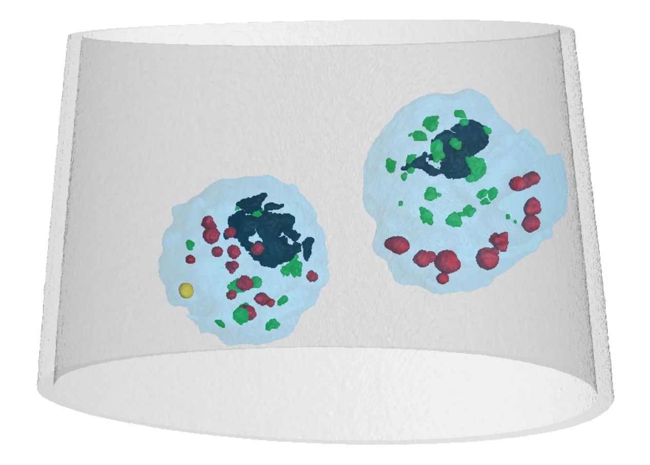
Biologists make use of a broad range of analytical tools to characterize the size, elemental composition and molecular structure of the different compartments inside the cell. Mass density measurements are also relevant, as they can provide information about the packing of the molecules in each compartment. Ideally such measurements should be done in situ, such that the location of each compartment remains in context with the entire cell or tissue in which it is embedded. However current density analytical tools such as density gradient centrifugation require the extraction of the compartments from the cell to be investigated.
Researchers at the Paul Scherrer Institute PSI have obtained a three-dimensional density map of intact Chlamydomonas unicellular algae using a novel technique called X-ray ptychographic computed tomography (XPCT). In order to fix the sample during the measurements, cells in their medium were rapidly frozen after confinement in a glass microcapillary and kept at cryogenic temperature during the entire experiment, which was conducted at the Swiss Light Source SLS. Unlike conventional absorption-based imaging techniques, phase imaging enabled by XPCT provides high sensitivity to the mass density of the specimen, allowing absolute density values to be directly obtained from the gray level tomographic reconstruction [1]. As a result, accurate density values were provided for certain organelles inside the cell such as starch granules or starch platelets around the pyrenoid.
Apart from providing absolute mass density maps, PXCT will enable 3D microscopy of extended frozen hydrated biological tissue on the nanoscale. In this proof of principle demonstration resolution was limited by thermal and mechanical stability to about 180 nm. However, PSI researchers expect that resolution can be improved drastically in the near future with the development of a new instrument with a dedicated cryo sample holder in vacuum with positioning accuracy on the nanometer scale [2,3].
References
[1] A. Diaz et al., Phys. Rev. B 85, 020104(R) (2012) DOI:10.1103/PhysRevB.85.020104[2] M. Holler et al., Rev. Sci. Instrum. 83, 3857 (2012) DOI:10.1063/1.4737624
[3] M. Holler et al., Sci. Rep. 4, 3857 (2014) DOI:10.1038/srep03857
Read the full story
Three-dimensional mass density mapping of cellular ultrastructure by ptychographic X-ray nanotomographyA. Diaz, B. Malkova, M. Holler, M. Guizar-Sicairos, E. Lima, V. Panneels, G. Pigino, A. G. Bittermann, L. Wettstein, T. Tomizaki, O. Bunk, G. Schertler, T. Ishikawa, R. Wepf, A. Menzel.
J. Struct. Biol. 192, 461-469 (2015) DOI:10.1016/j.jsb.2015.10.008
Contact
Dr. Ana Diaz, Swiss Light SourcePaul Scherrer Institut, 5232 Villigen PSI, Switzerland
Phone: +41 56 310 5626, e-mail: ana.diaz@psi.ch