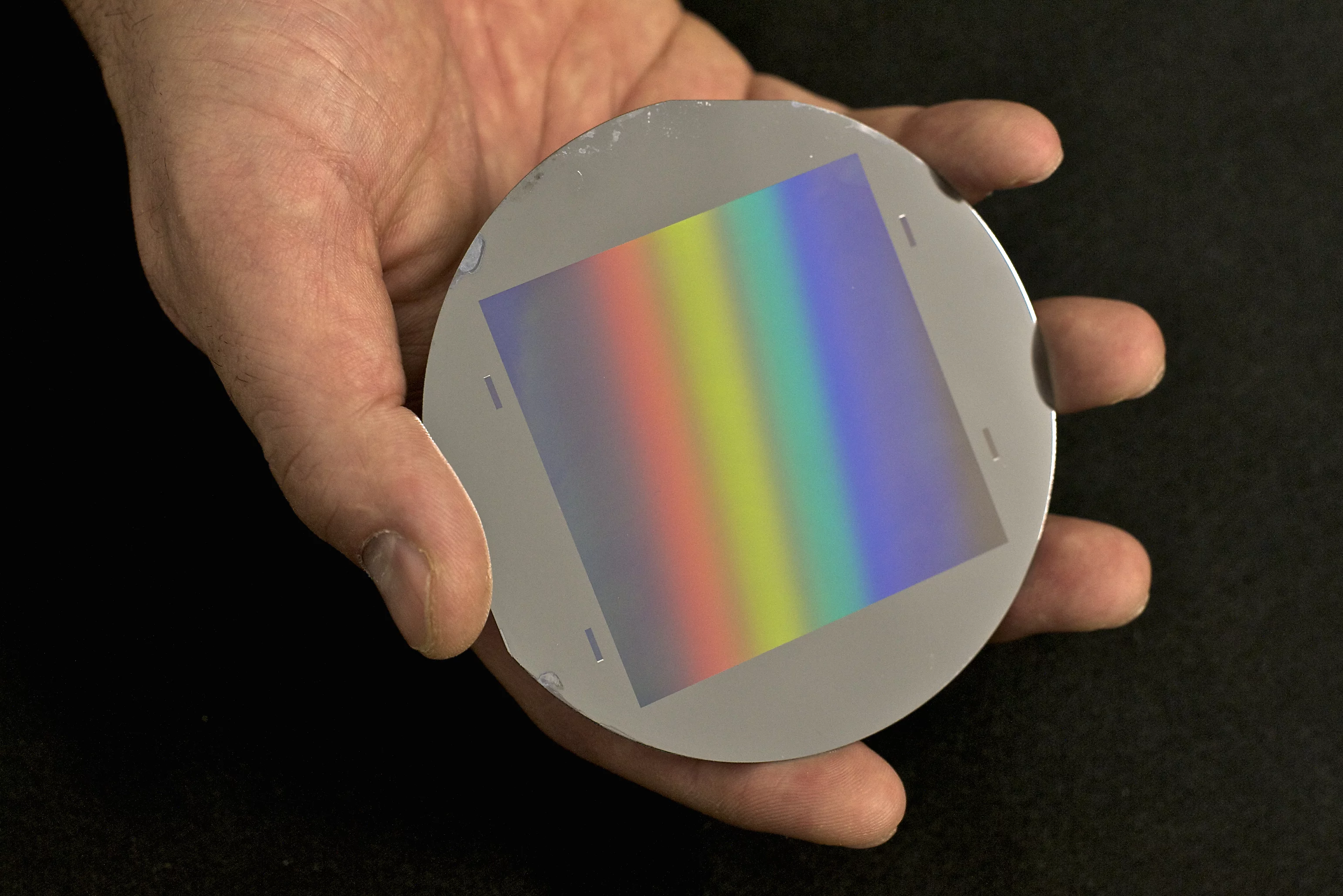
Figure 1: One of the gratings with fine lines in the micrometer range manufactured at PSI as used in the neutrons grating interferometer. The wafer has a diameter of 100 mm. The grid area is 64 x 64 mm2. The rainbow is caused by the refraction of light at the fine structures of the grating.
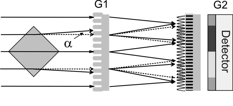
Figure 2: Schematical setup of neutron grating interferometer consisting of a phase and an absorption grating and an imaging detector. With the help of this setup neutrons can be detected, which are deflected or scattered at an angle of 10-4 degrees.
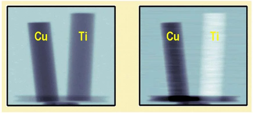
Figure 3: Measured neutron images of two metallic rods with 6 mm diameter made of copper and titanium. (Left) Conventional absorption image. (right) phase contrast image.
Phase contrast and dark-field images with visible light are indispensable tools for the modern microscopy technology. PSI had succeeded to develop the corresponding imaging techniques for neutrons. Hereby quantum mechanical interaction of neutrons with matter can be made visible in two-dimensional and three-dimensional images.
Particle physicists consider neutrons as small particle. Due to the wave-particle dualism neutrons can be described by matter waves, with a certain wave length. In difference to the conventional absorption contrast, where the contrast differences arise from the different attenuation of the materials, the image information in the phase contrast and dark-field images originate from the change of the wavelength in the material.
In the case of the phase contrast method one uses the fact, that waves which transverse an object having a different velocity than the waves which do not transverse the object. They therefore have a different wavelength. The therewith resulting displacement of the wave maxima leads to a change of the propagation direction and therefore to an angular change. An example from the classic geometrical optics clarifies how a phase sensitive image can be obtained. Considering a beam path through a collection lens, it follows from the law of refraction that initially parallel light rays are refracted towards the optical axis. In a wave-optical description this corresponds to lens induced angular change of the light rays, and namely a spatially dependent phase shift of the wave front.
In order to obtain phase contrast imaging, we measure the local angular variation of the neutron beam caused by the object. The experimental difficulty is, therefore, to measure efficiently for a variety of image points such small diffraction angles which are in the range of 10-4 degrees. Therefore one uses two grids (G1 and G2) which are composed of lines with lattice constants of a few micrometers. Such a grating is shown in Figure 1. The gratings together with a spatially resolving neutron detector then form the so-called neutron grating interferometer as depicted in Figure 2. The local angular change can be determined by analyzing pixel wise the measured intensity.
Results
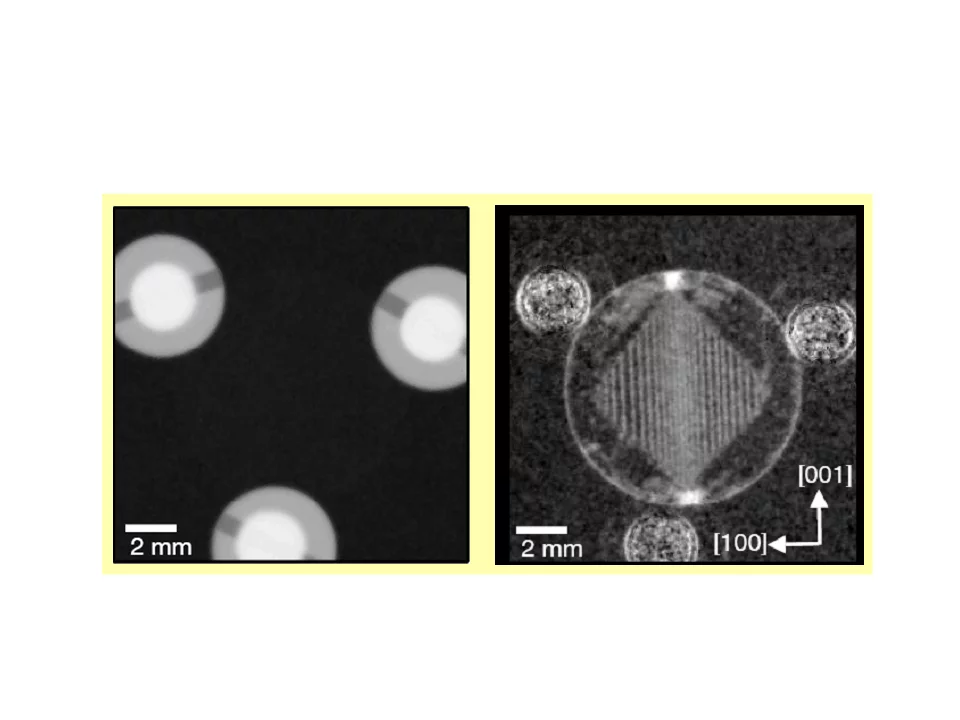
Figure 4: Measured neutron images of a magnetic sample (silicon iron) in the form of a disk with a thickness of 300 microns and 10 mm in diameter. (Left) Conventional absorption image. (right) Dark-field image. The white lines which form a rhombus are magnetic domain walls.
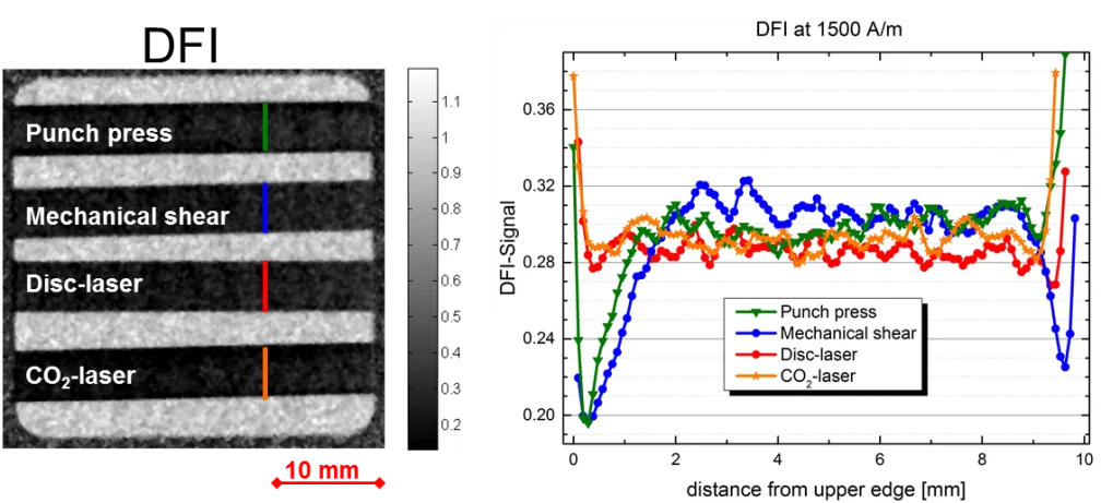
Figure 5: (Left) Dark-field image of 4 differently cut non-oriented FeSi electrical steel laminations. (Right) Profile along colored line. Mechanical cut samples show a decreased dark-field signal at the edge (punch press is only cut on the left hand side), while laser-cut sample don’t show this edge effect.
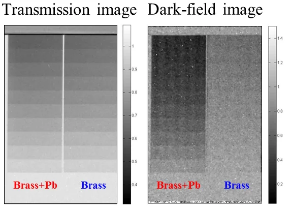
Figure 6: (Left) The transmission image of two brass step wedges. The left wedge is containing 3% of lead but no difference is reveal in this image. (Right) The dark-field image however clearly highlights the ability of detecting scattering, occurring from lead precipitations in the brass wedge.
Measurements of the phase shift of neutrons in interferometry experiments have been used so far to experimentally verify quantum mechanical predictions. The work at PSI aims to combine the available information about the phase shift with a space-resolved imaging technique, to image quantum mechanical interaction of neutrons with matter and to offer new contrast possibilities.
Figure 3 shows the results obtained for test samples. The conventional neutron absorption image (left) shows no measurable difference in the attenuation behavior of the two metals comprising of copper and titanium. The image of the measured phase shift (right), however, clearly shows a difference. Particularly interesting is the opposite sign of the phase shift of copper (black in the image against the gray background) and titanium (white to gray). This is a consequence of the different signs of the refractive indices of the materials.
For the dark-field imaging method the scattering properties in the in interior of materials is considered. Neutrons experience during the passage of the sample, which consist of materials with different refractive indices, as well a small angular change. This can be used for example to visualize magnetic domains in ferromagnetic materials, as shown in Figure 4. Neutrons are deflected due to the interaction of the neutron spin with the local magnetic fields, because magnetic domains with different orientations have different refractive indices. Therefore, neutrons are scattered in the transition from one domain to the next at the domain walls. The domain walls are seen in the dark-field image as white lines.
Another Example is the investigation of electrical steel sheet, used in nearly every type of electrical machines. The magnetic properties of electrical steel in magnetic cores such as stator and rotor laminations of electrical machines are known to be influenced by manufacturing conditions such as punching, interlocking or laser cutting. The effect of cutting is shown in figure 5. Two mechanical cutting techniques are compared with two laser cutting techniques. The mechanical techniques show a strongly reduced dark-field signal near the cutting edge, interpreted as a higher density of magnetic domain walls that is undesired and increases magnetic core losses. Furthermore it is not only magnetic scattering that can be detected like figure 6 shows. Two brass step wedges are shown here. The only difference is 3% of lead that is forming small precipitations in the left wedge. Since lead has a different refractive index for the traversing neutron the neutrons are scattered by the lead inclusions resulting in a measurable signal in the dark field image.
Publications
Visualizing the propagation of volume magnetization in bulk ferromagnetic materials by neutron grating interferometry.
C. Grünzweig, C. David, O. Bunk, J. Kohlbrecher, E. Lehmann, Y. W. Lai, R. Schäfer, S. Roth, P. Lejcek, J. Kopecek, and F. Pfeiffer
J. Appl. Phys. 107, 09D308 (2010).
DOI: 10.1063/1.3365373
Bulk magnetic domain structures visualized by neutron dark-field imaging.
C. Grünzweig, C. David, O. Bunk, M. Dierolf, G. Frei, G. Kühne, R. Schäfer, S. Pofahl, H. Rønnow, and F. Pfeiffer.
Appl. Phys. Lett. 93, 112504 (2008).
DOI: 10.1063/1.2975848
Neutron decoherence imaging for visualizing bulk magnetic domain structures.
C. Grünzweig, C. David, O. Bunk, M. Dierolf, G. Frei, G. Kühne, J. Kohlbrecher, R. Schäfer, P. Lejcek, H. Rønnow, and F. Pfeiffer
Phys. Rev. Lett. 101, 025504 (2008).
DOI: 10.1103/PhysRevLett.101.025504
Design, fabrication, and characterization of diffraction gratings for neutron phase contrast imaging.
C. Grünzweig, F. Pfeiffer, O. Bunk, T. Donath, G. Kühne, G. Frei, M. Dierolf, and C. David.
Rev. Sci. Inst. 79, 053703 (2008).
DOI: 10.1063/1.2930866


