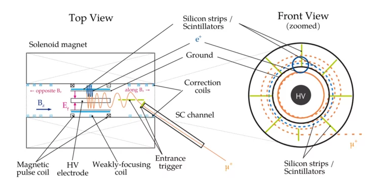
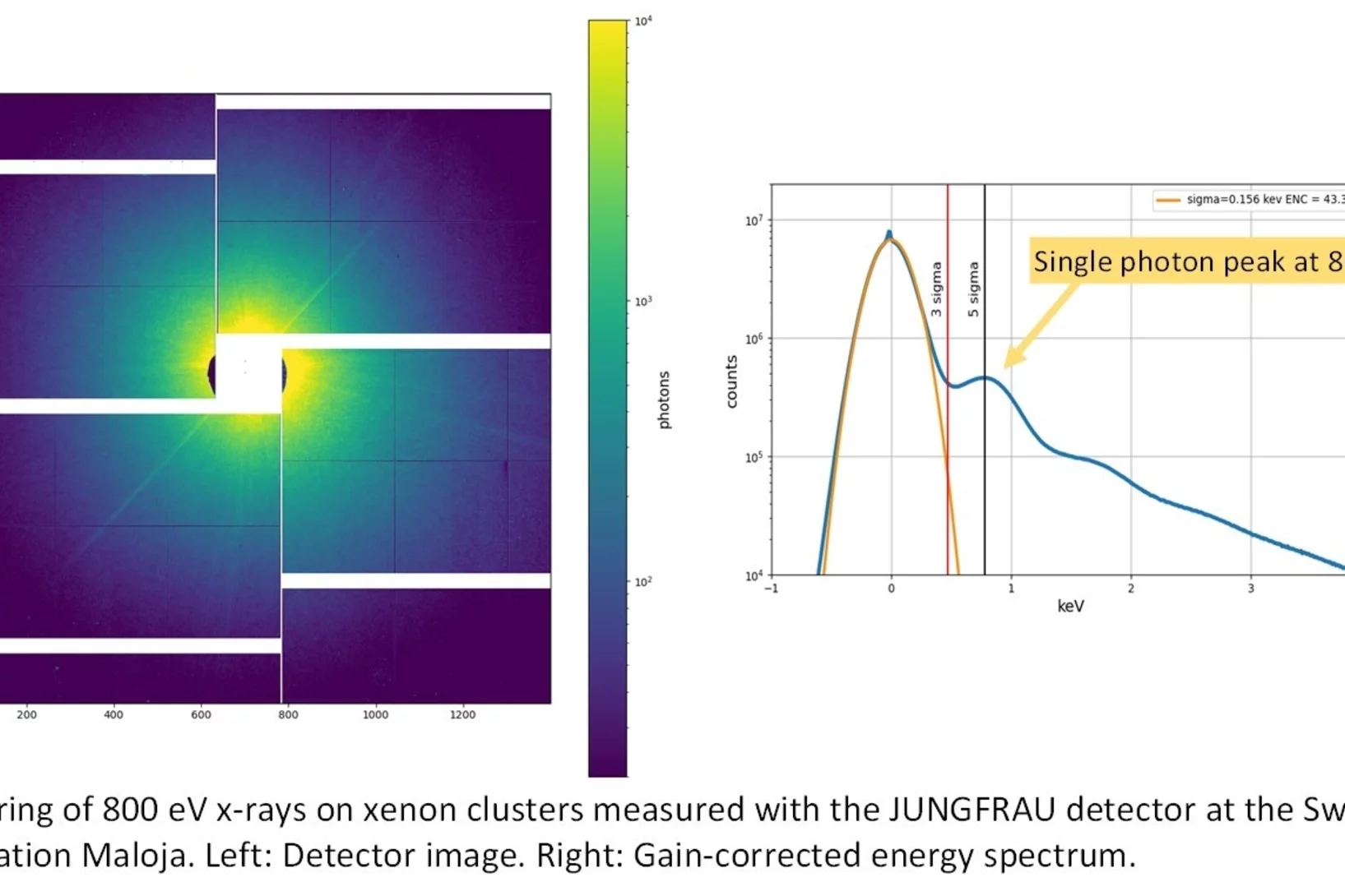
In the paper, the international muEDM collaboration at PSI discusses systematic effects of the most sensitive measurement of the muon's electric dipole moment (EDM). Scientists from Europe are developing a prototype experiment using the frozen-spin technique (FST) to achieve unprecedented sensitivity. The FST meticulously aligns a magnetic field with a perpendicular electric field so that the muon's spin orientation always follows its momentum. This enhances the sensitivity to the muon EDM by about 3 orders of magnitude compared to the best result from the muon g-2 experiment at Brookhaven National Lab.
The paper addresses systematic effects that could mimic an EDM signal when E- and B-fields are not perfectly aligned, adjusted, or stable over time. While most effects cancel out when reversing the magnetic field, some residual effects the specifications for the fields' uniformity, stability, and orientation,
which are challenging but achievable.