TOMCAT 2.0
The text here mirrors the report prepared by F. Marone, A. Bonnin, C. M. Schlepütz, G. Mikuljan, P. Zuppiger, and M. Stampanoni, and presented in the SLS 2.0 Beamline Conceptual Design Report (p. 353ff) published on January 25, 2021.
Click here for more info on the PSI Upgrade Project SLS 2.0.
In a nutshell
In the last decade, TOMCAT has been providing cutting-edge tomographic microscopy to a heterogeneous international scientific community and has generated seminal contributions in key areas like (bio-)medical research, material science, energy, geoscience, and paleontology, to cite just a few. Internationally, TOMCAT is considered a benchmark and is presently driving the developments of dynamical tomographic microscopy. To maintain and strengthen our leading role in the field, we plan to capitalise on our expertise in multiscale, multimodal dynamic tomographic microscopy and propose to deploy the TOMCAT 2.0 upgrade on two beamlines, dubbed I-TOMCAT, a brand-new instrument based on an insertion device of the latest generation, and S-TOMCAT, powered by a new high-field superconducting bending magnet. The embedment of SLS 2.0 within PSI will allow the implementation of innovative technical solutions in a very short time: at a pace that competitors will not be able to follow. TOMCAT 2.0 will be unique in offering one entry point for complex dynamical, high-throughput multidimensional imaging tasks requiring a spatial resolution ranging from 100 nm up to 10 μm and energies from 8 up to 80 keV. TOMCAT 2.0 on SLS 2.0 will provide images of increased quality, boosted by the smaller source size and higher photon flux at most energies. The upgrade will lead to better and novel characterization techniques, with simultaneously higher spatial and temporal resolution, pushing dynamic tomographic imaging towards unexplored frontiers. Extensive capabilities for in-situ, operando, in-vivo and in-fieri experiments will be offered to broad academic and industrial communities addressing grand challenges in medical, energy and the material sciences. The TOMCAT 2.0 upgrade will enduringly establish PSI on the map as one of the best imaging facilities worldwide for the next 20 years.
1. Upgrade motivation - goals - expected impact
X-ray tomographic microscopy provides quantitative volumetric information on a large variety of opaque samples in a fast and non-destructive manner with micrometer spatial resolution for samples of a few millimetres in diameter. The high brightness of synchrotron radiation sparked the development of multi-modal imaging techniques, including phase contrast, dark-field imaging, and diffraction CT. Over the last few years, new fast camera and readout systems [1] have triggered the advance of dynamic tomographic studies, to a point where processes can be followed in 3D up to speeds of 20 tomograms (i.e., 3D volumes) per second at a few microns spatial resolution. TOMCAT has been at the forefront from the very early days of synchrotron-based tomography, both for fast tomographic measurements and for multi-modal imaging. In the last decade, TOMCAT has been providing cutting-edge tomographic microscopy in different flavours to a heterogeneous international scientific community and has generated seminal contributions in key areas like (bio-)medical research [2], material science [3], energy [4], geoscience [5], and paleontology [6], to cite but a few.
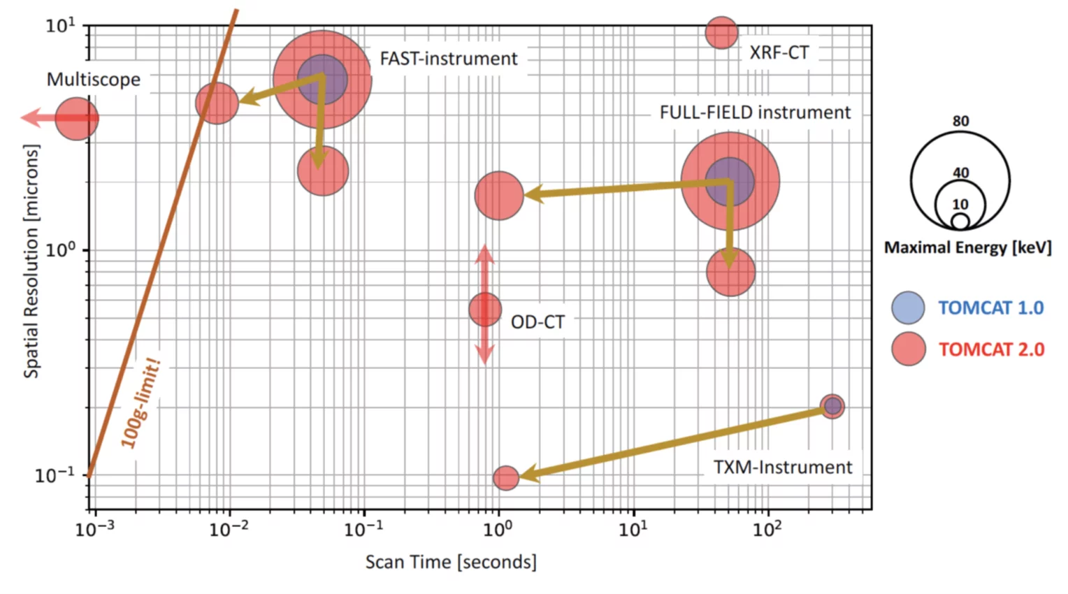
In addition to a significant overall increase in image quality boosted by the higher photon flux at most energies, the unprecedented beam properties of SLS 2.0 will lead to better and novel characterization techniques, with simultaneously higher spatial and temporal resolution, pushing dynamic tomographic imaging towards unexplored frontiers. We plan to capitalize on our expertise in multiscale, multimodal, dynamic tomographic microscopy and propose to deploy the TOMCAT 2.0 upgrade at two beamlines, dubbed I-TOMCAT a brand-new instrument based on an insertion device of the latest generation, and S-TOMCAT powered by a high-field superconducting bending magnet. With two beamlines, TOMCAT 2.0 will offer a unique and comprehensive imaging portfolio at energies ranging from 8 up to 80 keV. Figure 1 illustrates the expected improvement in tomographic capabilities at TOMCAT 2.0 compared to the present status. The figure shows the time needed to acquire a 3D volume at a given 3D resolution for different instruments and techniques.
TOMCAT 2.0 will enable the study of dynamic processes over an unparalleled length scale range (100 nm up to 10 μm), to some extent even in a simultaneous manner, an important aspect for processes with hierarchical structures. We foresee up to three orders of magnitude increase in time resolution for the Transmission X-ray Microscopy (TXM) instrument with a target spatial resolution of at least 100 nm and almost a doubling of the operating energy range. The full-field instrument will experience almost two orders of magnitude improvement in speed and up to one order of magnitude better spatial resolution for the same energy. For the same time and spatial resolution, it will be possible to double (!) the probing energy, enabling the investigation of significantly thicker and heavier samples compared to now. Similar arguments hold for the fast instrument. Its time-resolution will be pushed to the physical barrier imposed by centrifugal forces acting during sample rotation. Overcoming this barrier means entering the domain of single-shot kHz (and above) 3D imaging, which will require illuminating the sample simultaneously from multiple directions under limited — or eventually even no — specimen rotation. Time resolutions in the kHz regime have been achieved already thanks to post-gating techniques.
The higher flux and larger fraction of coherent photons at higher energies as well as a palette of specialized in-situ, operando, in-vivo, and in- fieri sample environments will be instrumental for the investigation of dynamic phenomena under realistic conditions (e.g., less boundary effects) and will expand the scope of current high-speed capabilities to new systems and scientific questions. Low-dose in-vivo studies on small and larger organisms will also strongly benefit from the unrivalled beam properties at SLS 2.0. The investigation of large volumes at high resolution — stitched teravoxel tomography [7] — will become routine. TOMCAT 2.0 dynamic aspects will be complemented by 3D multimodal capabilities revealing, in addition to the 3D microstructural evolution, information on the elemental distribution (full-field x-ray fluorescence tomography), crystallographic orientations (white-beam diffraction contrast tomography) and structural anisotropy (omnidirectional Talbot interferometry).
1.1 Uniqueness compared to other present and planned beamlines worldwide
Essentially all synchrotron facilities worldwide operate or plan a hard x-ray TXM or a full-field tomography beamline. Consequently, both new and currently upgrading facilities all have a strong imaging program in their refurbishment portfolio. TOMCAT is internationally considered a benchmark; it has established itself as a world reference for tomographic imaging performed at energies between 8 and 30 keV, making very good use of the excellent performances of the SLS machine in that particular energy range. The documented great success of the beamline with more than 600 papers published so far and a ten-years average overbooking factor of 1.6 — and this, despite data acquisition becoming more than ten times faster during the last few years — clearly places TOMCAT at the forefront of the synchrotron imaging community. Several facilities have plans to build imaging beamlines inspired by TOMCAT (AS in Melbourne, PAL in Pohang, SSRF in Shanghai, ALBA in Barcelona, HEPS in Beijing, MOGNO in Campinas, BEATS in Amman), while others have clearly identified (full-field) imaging as their ”raison d’ ˆetre” for their upgrade programs (ESRF and APS in particular).
The proposed TOMCAT 2.0 upgrade foresees the implementation of a number of high-potential technical innovations which will guarantee us a leading position in the imaging community for the next two decades. We are not aware of any other institution able to design, construct, install, and commission a superconducting short-period undulator and a superconducting high-field longitudinally graded bending magnet, while simultaneously offering access to resources conceiving and fabricating both novel optics and detectors: such a synergetic effort is only possible at PSI and will enable an efficient implementation of innovative ideas at a pace that competitors will not be able to keep. No one worldwide can/will offer such a flexible operative range as TOMCAT 2.0 will be able to do, as most of the competitors usually run full-field and TXM endstations with independent, often not even collaborating, teams. Our crew will share knowledge and distribute it on either I-TOMCAT or S-TOMCAT ensuring the latest developments and protocols are implemented and exploited.
1.2 Impact of the new ring brilliance
At SLS 2.0, we expect a significantly larger horizontal transverse coherence length compared to SLS today, given by the much-improved horizontal emittance properties of the SLS 2.0 machine. This will lead to an almost isotropic transverse coherence function — for S-TOMCAT the source actually becomes slightly smaller and round, while now we have roughly a 7:1 (H:V) ratio — which will translate into a generally improved image quality for all phase-contrast techniques implemented at TOMCAT 2.0: propagation-based, grating interferometry, Zernike and, in the long term, single-shot. In its simplest form, phase-contrast imaging relies on the detection of interference fringes arising from a subtle refraction of the x-ray beam at interfaces between materials with slightly different refractive indices. In the current SLS, the horizontal fringe visibility is significantly reduced due to source size blurring, an effect which is exacerbated by increasing the propagation distance (and hence reducing the source demagnification ratio of the imaging system). Much sharper fringe patterns — resulting from the increased SLS 2.0 brilliance — will dramatically boost phase-contrast sensitivity. New phase reconstruction algorithms to take optimal advantage of that situation are already under development in the community [8]. This will be particularly important for coarse resolution scans in multiscale imaging and high-energy experiments, both of which require larger propagation distances; and for high-speed measurements, where the enhanced fringe visibility will allow feature detection even at very low signal to noise ratios.
It has to be pointed out that not only phase-sensitive techniques will profit from the improved ring brilliance. One advantage of SLS 2.0 (compared to other – larger – rings) is that due to its small dimension it is actually possible to install optical components very close to the source. For a 2 m short-straight undulator and a predicted machine emittance reduction of 30x along the horizontal direction, we expect a significant reduction of horizontal source size and divergence compared to TOMCAT. We calculated that a refractive axicon [9] placed at 15 m from the source will collect the full undulator beam with a transmission efficiency higher than 97% at 12 keV. This will allow us to efficiently operate the TXM at 400x magnification with a working distance of 50 mm. In combination with a MOENCH detector, this will result in a pixel size of 65 nm and we expect to collect about 4000 photons/pixel/frame at 1000 frames/s. The present MOENCH0.3 design can handle a maximum of 1000 photons/pixel/frame. Future designs might perform better in this respect and will further contribute towards better image quality. This means an improvement in speed of about 2000x compared to what we can do now on TOMCAT, see Figure 1.
1.3 Complementarity to other PSI BLs
TOMCAT 2.0 will focus mainly on the dynamic multi-modal aspects of tomographic microscopy. Our goal will be to provide time-resolved image volumes of evolving samples, in situ, in vivo, operando, or in fieri. We target a maximum spatial resolution of 100 nm, while chemical discrimination will be in the order of 600 to 900 eV. In this sense, TOMCAT 2.0 will be complementary to cSAXS (where spatial resolution is pushed to the physical limits) and microXAS (where chemical imaging is the daily business and can be performed at higher spatial resolution). We are and will continue cooperating with the cSAXS and microXAS teams, for instance in the development of ptychographic methods as well as full-field XRF-CT and WB-DCT. Other beamlines are planning to reinforce their imaging portfolio (Phoenix, PolLux, and SIM in particular) and we will be very happy to share our knowledge in tomographic imaging with them.
1.4 Size and impact of community – potential increase through the upgrade
TOMCAT’s 10 years average overbooking factor is 1.6, while during the last five years, it has risen to 1.8. These can be considered healthy numbers and they confirm a sustained interest in the beamline, which has been fully booked for more than a decade. In view of the SLS 2.0 upgrade, we organized a workshop in November 2016, where we invited key opinion leaders (KOLs) from our broad user community to brainstorm on the mission of TOMCAT 2.0. Physical limitations of the current TOMCAT beamline were discussed and game-changers as well as new science enablers were identified in:
- the capability to dynamically resolve 3D morphological details down to 100 nm (in situ, in fieri, operando, in vivo);
- the ability to visualize sub-micron functional features (like fiber orientation in a polymer material) on large areas;
- the implementation of correlative microscopy options (fluorescence information on top of structural details).
These are features which can only be successfully implemented on an undulator beamline, as proposed for I-TOMCAT. Last, but not least, imaging up to 80 keV has been identified as a key functionality to extend significantly TOMCAT 2.0 capabilities and its user portfolio. This feature can only be provided on SLS 2.0 by using a high-field superconducting bending magnet, as suggested for S-TOMCAT. The proposed conceptual design for TOMCAT 2.0 has thus been driven by the imaging needs dictated by the science carried out by world-leading scientists. A letter of support in this sense signed by 12 KOLs was attached to the November 2016 report. With a real-time hard x-ray TXM operated synergistically with a high-energy full-field imaging setup, TOMCAT 2.0 will capitalize on a decade of cutting-edge tomographic microscopy expertise and attract a plethora of new users fully exploiting the new opportunities.
1.5 Industrial potential
TOMCAT is being regularly booked by industrial customers and their interest in multiscale, dynamical tomographic imaging is constantly growing. Our proprietary research reflects the flexibility of the TOMCAT instrument and our industrial requests come from very different areas (like pharmaceutical, energy or space technology). We do not offer a standardized product, but rather a customized service to target specific problems. We advise customers on how to design their synchrotron experiment and we help them during the beamtime and whenever possible in the quantification of the generated data: this comprehensive service package is a very much appreciated asset. A successful industrial beamtime requires a significant effort from the local team, before, during and after a proprietary beamtime. In March 2019, we hired an industrial liaison scientist, dedicated to this task and with the charge to develop an even more complete and competitive industrial program on TOMCAT 2.0. The flexibility of our technology is mirrored by the heterogeneity of our industrial community. Within the context of a EU-funded project proposal, we recently asked for Letters of Support (LOSs) and the response from industry was enthusiastic, with supportive statements from ABB, Novartis, Toyota, Shell, TyssenKrupp, Lument and SAFTd. Although it might be difficult to group all those companies into a consortium willing to co-fund a complete beamline, we think that such a strong interest from a broad variety of companies is actually a very healthy operational context as our industrial income is diversified and hence does not depend on a single industrial area.
References
1. R. Mokso, C. M. Schlepütz, G. Theidel, H. Billich, E. Schmid, T. Celcer, G. Mikuljan, L. Sala, F. Marone, N. Schlumpf, and M. Stampanoni. Gigafrost: the gigabit fast readout system for tomography. Journal of Synchrotron Radiation, 24:1250–1259, 2017.
2. A. Lathuiliere, V. Laversenne, A. Astolfo, E. Kopetzki, H. Jacobsen, M. Stampanoni, B. Bohrmann, B. L. Schneider, and P. Aebischer. A subcutaneous cellular implant for passive immunization against amyloid-beta reduces brain amyloid and tau pathologies. Brain, 139:1587–1604, 2016.
3. L. K. Aagesen, A. E. Johnson, J. L. Fife, P. W. Voorhees, M. J. Miksis, S. O. Poulsen, E. M. Lauridsen, F. Marone, and M. Stampanoni. Universality and self-similarity in pinch-off of rods by bulk di↵usion. Nature Physics, 6(10):796–800, 2010.
4. Patrick Pietsch, Daniel Westhoff, Julian Feinauer, Jens Eller, Federica Marone, Marco Stampanoni, Volker Schmidt, and Vanessa Wood. Quantifying microstructural dynamics and electrochemical activity of graphite and silicon-graphite lithium ion battery anodes. Nature Communications, 7:12909, 2016.
5. D. R. Baker, F. Brun, C. O’Shaughnessy, L. Mancini, J. L. Fife, and M. Rivers. A four-dimensional X-ray tomographic microscopy study of bubble growth in basaltic foam. Nature Communications, 3, 2012.
6. D. J. E. Murdock, X. P. Dong, J. E. Repetski, F. Marone, M. Stampanoni, and P. C. J. Donoghue. The origin of conodonts and of vertebrate mineralized skeletons. Nature, 502(7472):546, 2013.
7. A. Miettinen, A Bonnin, A. Patera, F. Marone, and Stampanoni M. Non-rigid stitching of teravoxel-scale volumetric images. Bioinformatics, 2019.
8. J. Villanova, R. Daudin, P. Lhuissier, D. Jauffres, S. Y. Lou, C. L. Martin, S. Laboure, R. Tucoulou, G. Martinez-Criado, and L. Salvo. Fast in situ 3D nanoimaging: a new tool for dynamic characterization in materials science. Materials Today, 20(7):354–359, 2017.
9. D. Zverev, A. Barannikov, I. Snigireva, and A. Snigirev. X-ray refractive parabolic axicon lens. Optics Express, 25(23):28469–28477, 2017.