Research Archive

Here is a summary of some of the previous research activities within the TOMCAT team. Click on an item to be directed to a more detailed project description.

Dynamic in-vivo lung imaging at the micrometer scale

In vivo tomographic slice of a newborn rat’s lung with alveolar resolution.
With the recent development and realization of in vivo tomographic X-ray microscopy for the study of lung physiology at the micrometer scale [1] we are now able to study a variety of phenomena that take place in the mammalian lung, or more precisely, in a murine and rat animal model. Following that, we are currently pursuing two parallel lines of research. On the one hand, we are constantly improving our established imaging technique in terms of acquisition speeds, dose reduction and post-processing. Here we follow the ultimate goal towards a free-breathing microscopic lung imaging animal model, but the methods remain transferable to other in vivo studies as well. On the other hand, we have now the availability to study for the first time the detailed (microscopic) mechanisms behind various lung diseases. As the first example, we are currently investigating the origins of ventilator-induced lung injury (VILI), which is being one of the major reasons for the high mortality in ventilator-treated patients with acute respiratory distress syndrome (ADRS) – a pathological condition where ventilator treatment is highly necessary.

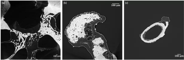
Apart from that, our interests also extend to the study of other lung diseases such as emphysema and fibrosis. Our results will have direct implications on the current knowledge and understanding when dealing with lung diseases in clinics. For instance, a fundamental understanding of VILI would be crucial for developing better ventilation strategies for patients.

Publications:

  1. G. Lovric, ETH Phd thesis, 2015, http://doi.org/bd3z.
  2. G. Lovric, S. F. Barré, J. C. Schittny et al., J. Appl. Crystallogr. 46 (4), 856, 2013, http://doi.org/n3p.

Collaboration:

  • Institute of Anatomy, University of Bern, Switzerland
  • Clinic of Neonatology, University Hospital of Lausanne (CHUV), Switzerland
  • Department of Clinical Physiology, Grenoble University Hospital, France
  • Department of Surgical Sciences, Uppsala University, Sweden

Funding agencies: Centre d'Imagerie BioMédicale (CIBM)

X-ray phase contrast tomographic imaging and analysis of the lung at the micrometer scale

Phase contrast tomographic reconstructions of intact mouse lungs at the micrometer scale.
The gas exchange between the ambient air and the blood takes place in the mammalian lung. The area at the distal part of the organ is subdivided into functional units called acini, which start distally of the terminal bronchioles. Despite of their importance, limited knowledge exists about their three-dimensional (3D) dynamics and development, as the borders between acini are not detectable in classical two-dimensional (2D) histology slices. Furthermore the histological fixation makes any kind of dynamical studies infeasible. Recent developments in synchrotron based X-ray tomographic microscopy helped us to overcome these limitations. Volumetric lung datasets of fresh post-mortem murine lungs with micrometer spatial and sub-second temporal resolutions at different pressure levels are nowadays routinely acquired using synchrotron propagation based phase contrast X-ray tomographic microscopy at the TOMACT beamline of the Swiss Light Source [1]. The aim of this project is to investigate the 3D structural changes of the acinus at the micrometer scale at different pressure levels and days of lung development. Due to the complexity of the lung microstructure and the size of individual acini a specialized 3D segmentation algorithm [2], as well as a technique to enlarge the field of view has been developed [3]. Our methods can run in parallel on multiple datasets. By analyzing a large number of samples we aim to extract the acinar skeleton and address the question of the acinar deformation patterns by quantifying local structural differences at different pressure levels with statistical significance.

Publications:

  1. Lovric, Goran, et al. "A multi-purpose imaging endstation for high-resolution micrometer-scaled sub-second tomography." Physica Medica (2016), http://doi.org/bp54.
  2. Vogiatzis Oikonomidis Ioannis et al. "Efficient segmentation of lung parenchyma in tomographic images of freshly post mortem mice under low contrast-to-noise ratio conditions and at micrometer resolution. " XRM 2016 conference proceedings
  3. Vogiatzis Oikonomidis Ioannis et al. "Imaging samples larger than the field of view: the SLS experience." XRM 2016 conference proceedings

Collaboration:

  • Institute of Anatomy, University of Bern, Switzerland

Funding agencies: Swiss National Science Foundation (SNF)
 

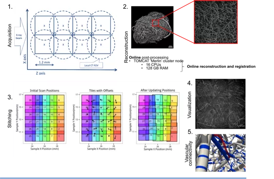
Reconstruction of the mouse brain vascular networks with high-resolution synchrotron radiation X-ray tomographic microscopy

A schematic overview of the project: from image acquisition to processing of TB-sized dataset of the mouse brain.
Brain vessels play an important role in the process of maintaining normal brain function. An in-depth knowledge of the vascular structure and topology is essential for better understanding the pathophysiological cerebral processes. Within the context of the Human Brain Project (HBP), this project aims to reconstruct, in a non-destructive way, the entire vascular system of the mouse brain with high-resolution. Synchrotron-radiation X-ray phase-contrast tomographic microscopy at the Swiss Light Source of the Paul Scherrer Institute (Switzerland) is used as a non-invasive key technology for fast image acquisition of mouse brain samples, previously perfused with contrast agent and kept in steady-state conditions. Current sample preparation procedure suggests an optimal perfusion procedure based on indian ink [1]. However, new sample preparation methods are explored within the project. To fully reconstruct the complete cerebrovascular network of the mouse brain with 1μm resolution, several local tomographic scans need to be acquired, reconstructed and stitched together [2] in order to cover the whole brain volume, thus leading to TB-sized datasets. All the pioneering efforts to address the challenging task of analysing such large datasets are pointing towards new horizons in the investigation of large biological samples with 3D high spatial resolution.

Publications:

[1] Xue S, Gong H, Jiang T, Luo W, Meng Y, et al. Indian-Ink Perfusion Based Method for Reconstructing Continuous Vascular Networks in Whole Mouse Brain. PLoS ONE 9(1): e88067 (2014), http://dx.doi.org/10.1371/journal.pone.0088067.

[2] Preibisch, S, Stephan S, Tomancak P. Globally optimal stitching of tiled 3D microscopic image acquisitions. Bioinformatics (Oxford, England) 25 (11): 1463–5 (2009), https://doi.org/10.1093/bioinformatics/btp184.

Collaboration:

  • University of Zurich, Institute of Pharmacology and Toxicology, Switzerland
  • IBFM - Inst. of Molecular Bioimaging and Physiology, Dept. of Biomedical Sciences, LITA Segrate (Milano), Italy
  • European Synchrotron Radiation Facility – ESRF Grenoble, Medical Beamline, France

Funding agencies: Centre d'Imagerie BioMédicale (CIBM)

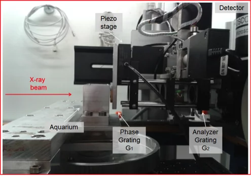
X-ray grating interferometry for phase-contrast imaging at the Swiss Light Source

DPC setup at the TOMCAT beamline of the Swiss Light Source.
With the development and realization of differential phase contrast (DPC) imaging based on grating interferometry (GI) [1], which provides high sensitivity to electron-density variations within soft tissues, we are now able to study a variety of phenomena in the medical field such as the amyloid plaque distribution in mouse brains [2] and the evolution of 3D substructures developing during the early stage of tumour formation [3]. Most recently, data acquisition and post-processing have been optimised at TOMCAT, thus enabling a full phase volume to be acquired in 32 min. As detector, a sCMOS camera with a 16-bit nominal dynamic range is attached to a 1:1 optical microscope which results in a 6.5 μm pixel size and 1.3 cm FOV for imaging. Our research is currently focused on the further improvements of the DPC setup in terms of acquisition speeds, dose reduction and post-processing. In addition to that, the ongoing efforts aim to improve the hard components (new gratings manufacture and setup versatility). On the other hand, we are also interested to experimentally exploit the capabilities of DPC imaging in a wider range of applications from medicine to material science specifically using synchrotron light.

Publications:

[1] McDonald SA et al. Advanced phase-contrast imaging using a grating interferometer. J. Synchr. Radiat. 16, 562 (2009), https://doi.org/10.1107/S0909049509017920.

[2] Pinzer B. R. et al. Imaging brain amyloid deposition using grating-based differential phase contrast tomography.Neuroimage 61, 1336-1346 (2012), http://dx.doi.org/10.1016/j.neuroimage.2012.03.029.

[3] Beheshti A. et al. Early Tumor Development Captured Through Nondestructive, High Resolution Differential Phase Contrast X-ray Imaging. Radiat, Res. 180, 448-454 (2013), http://dx.doi.org/10.1667/RR13327.1.

Funding agencies: Centre d'Imagerie BioMédicale (CIBM)

Impact of phase-contrast X-ray imaging in cochlear micro-anatomy investigation

Tomographic slices of: (a) ROI in the cochlea, (b) incus and (c) stapes. Images acquired at the TOMCAT beamline, PSI Switzerland.
The human cochlea is composed of about two and three-fourth turns, but unusual anatomy with cochlear three turns has been described [1]. It is surrounded by a compact bony structure and represents the hardest bone in the body with a trilamellar arrangement with islands of modified cartilage and high-mineral content, which increases the stiffness of the bony labyrinth [2]. There is now much interest on cochlear anatomy due to surgical approaches that electrically stimulate the auditory nerve (cochlear implantation, CI). In cochlear implantation (CI), the large variations in cochlear lengths, angles between turns, and position in the skull base can influence the straightforwardness for the insertion of a CI electrode particularly passing the first turn. Should we use personalized electrodes in such a way that they minimize trauma during insertion? This is important, because trauma against the medial wall of the ST (scala tympani) can damage the spiral ganglion, which is the prime target of electric stimulation by the electrodes of the implant. While a state of the art micro CT imaging device such as the Scanco μCT 100 can provide resolutions of less than 4 μm to answer the above-stated questions, its ability to image soft tissue is limited. For this reason, 1 μm resolution images of human cochlea with and without implants and ear ossicles will be acquired with synchrotron-based tomographic microscopy at the Swiss Light Source (SLS) of the Paul Scherrer Institut (Switzerland). This project aims to optimize imaging of bony and soft tissue of the middle and inner ear anatomy and characterize human cochlear micro-anatomy, by integrating it fully into a multimodal microatlas. This investigation will provide insightful information on morphology of normal cochlea anatomy and its variations before and after CI, permitting the study of cochlear trauma after CI and the creation of a multimodal microatlas of the cochlea.

Publications:

[1] Tian Q, Linthicum FH, Jr., Fayad JN: Human cochleae with three turns: an unreported malformation. Laryngoscope, 116(5):800-803 (2006), http://dx.doi.org/10.1097/01.mlg.0000209097.95444.59.

[2] Rask-Andersen H, Liu W, Erixon E, Kinnefors A, Pfaller K, Schrott-Fischer A, Glueckert R: Human cochlea: anatomical characteristics and their relevance for cochlear implantation. Anat Rec (Hoboken), 295(11):1791-1811 (2012), https://doi.org/10.1002/ar.22599.

Collaboration:

  • University of Bern, ARTORG Center for Biomedical Engineering, Switzerland
  • Inselspital Bern, Department of Otorhinolaryngology (ENT), Switzerland
  • PSI, Center for Proton Therapy, Switzerland

Funding agencies: Centre d'Imagerie BioMédicale (CIBM)

Virtual Reading of a Large Ancient Handwritten Science Book

Several examples of tomography reconstructed inner page portions revealing words and sentences (top), compared to visible pictures (bottom) [4].
The aim of this project is to implement a new X-ray tomography ”virtual reading” technique in order to read inside a large ancient handwritten book without opening it. The development of this technique is primarily inspired by the Venice Time Machine (VTM) project (EPFL, 2015). This is an ongoing collaboration between the Ecole Polytechnique Fédérale de Lausanne (EPFL) and two institutions in Venice: the University Ca’ Foscari and the ”Archivio di Stato”. The Archivio is an historical collection containing almost 100 kilometers of handwritten documents covering ten centuries of the administrative and legal life of Venice. But, as for all ancient collections, their exploitation by scholars is problematic for conservation and logistic reasons: without massive digitization, deciphering, indexing and storage, they are almost unusable. Within the context of a digitalization, X-ray imaging is used to analyze specimen without opening them [1, 2]. Tomographic reading is feasible thanks to the iron present in ancient inks (iron gall) over one millennium – whereas carbon or organic inks do not provide sufficient x-ray contrast. For this reason, a phase approache is also explored [3, 4].

Publications:

[1] Baumann, R., Porter, D. C., Seales, W. B. The use of micro-CT in the study of archaeological artifacts. Proc. of 9th Int Conf on NDT of Art, 1–9 (2008), http://citeseerx.ist.psu.edu/viewdoc/download?doi=10.1.1.216.2327&rep=rep1&type=pdf.

[2] Seales, W., Griffioen, W., Baumann, R., Field, M. Analysis of herculaneum papyri with x-ray computed tomography. Proceedings of 10th International Conference on NDT of Art, Jerusalem, 1–9 (2011), http://www.ndt.net/article/art2011/papers/FIELD%20-%20M%2014.pdf.

[3] Margaritondo, G. Elements of Synchrotron Light for Biology, Chemistry, and Medical Research. New York: Oxford (2002).

[4] Albertin F., Patera A., Jerjen I., Hartmann S., Peccenini E., Kaplan F., Stampanoni M., Kaufmann R., Margaritondo G., Virtual reading of a large ancient handwritten science book, Microchemical Journal, Volume 125, Pages 185-189, ISSN 0026-265X (2016), http://dx.doi.org/10.1016/j.microc.2015.11.024.

Collaboration:

  • EMPA, Center for X-ray Analytics, Switzerland
  • CIBM, Phase contrast X-ray imaging core (EPFL), Switzerland
  • Faculté des Sciences de Base, Ecole Polytechnique Fédérale de Lausanne (EPFL), Switzerland

Funding agencies: Centre d'Imagerie BioMédicale (CIBM)

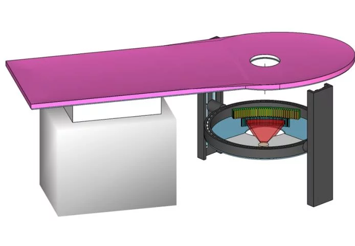
Full-Field Transmission X-ray Microscopy using a Photon Counting Pixel Detector

X-ray image of Star pattern made of gold acquired using the full-field transmission X-ray microscope with the Merlin detector (515 x 515 pixels of 55 microns). The photon energy was set to 7.5 keV to achieve an X-ray magnification of roughly 725x and effective pixel size of 76 nm.
Full-field transmission X-ray microscopy (TXM) can be used to investigate many materials and systems in biology, material science and energy sciences [1—3]. On the one hand, the performance of the TXM is determined by the quality of the X-ray optics. In particular, the outermost zone width of a Fresnel zone plate (FZP) used as objective lens determines the spatial resolution and its diffraction efficiency usually limits the speed of image acquisition. On the other hand, the spatially resolving detector used to acquire the magnified image of the sample is also a key element of the TXM system. To date, they usually consist of combination of a scintillating material and a visible, high numerical aperture microscope to collect the fluorescent visible light produced by the impinging X-ray photons. From the point of view of photon detection efficiency, low noise and high dynamic range, the use of single photon counting pixel detectors would be highly convenient. However, due to their large pixel sizes (> 55 μm) and the only moderate X-ray magnification (100x—300x) of the current TXM systems, the use of single photon counting pixel detectors has not been broadly considered. Within this project, we aim to combine the optimum diffractive X-ray optics with the long experimental hutches available to achieve large X-ray magnifications and make use of the advantages of photon counting pixel detectors.

Publications:

  1. A. Sakdinawat and D. Attwood. Nature Photonics 4, 840—848 (2010)
  2. M. Holt et al. Annu. Rev. Mater. Res. 43, 183—211 (2013)
  3. J. Vila-Comamala et al. J. Synchrotron Rad. 19, 705—709 (2012) DOI

Collaboration:

  • Dr. A. Parsons, Dr. E. Gimenez-Navarro and Dr. U. Wagner, Diamond Light Source, Didcot (UK)
  • Dr. C. David, Paul Scherrer Institut, PSI Villigen (Switzerland)

Funding agencies: FNSNF Grant Number 159263

Lossy Compression for CT Datasets

A dataset for tomographic reconstruction as obtained in laboratory setups typically consists of several hundred projective X-ray images of considerable size, quickly amounting to dozens or hundreds of gigabytes per experiment. For different reasons, among them archiving, it is desirable to keep the originals. Their transfer and storage impose a substantial – also financial – burden on the IT infrastructure, making it highly desirable to reduce the size of the files. Unfortunately, previous studies have shown that lossless compression has only limited impact due to hardly compressible image noise. On the other hand, lossy compression has long since become standard for many other imaging applications such as geographic information systems, trading some lost information for better compressibility.

The core idea of lossy compression is to subtly modify an image such that it can be compressed well while keeping the visual appearance as much as possible. For average compression ratios, this will typically lead to tiny ‘ghost’ structures around more prominent image features or smoothing that the human eye will miss. In the case at hand, however, where the images serve as input to the first stage of a longer processing pipeline, such modifications may lead to artefacts in the final tomographic reconstructions. This project aims at evaluating different lossy compression schemes and at investigating the impact of their respective artefacts onto reconstruction quality.

Publications:

  1. Impact of lossy compression of X-ray projections onto reconstructed tomographic slices, F. Marone, J. Vogel, M. Stampanoni, Journal of Synchrotron Radiation 27 (2020).

Contact: Dr. Federica Marone, federica.marone@psi.ch, WBBA/216, +41 56 310 53 18

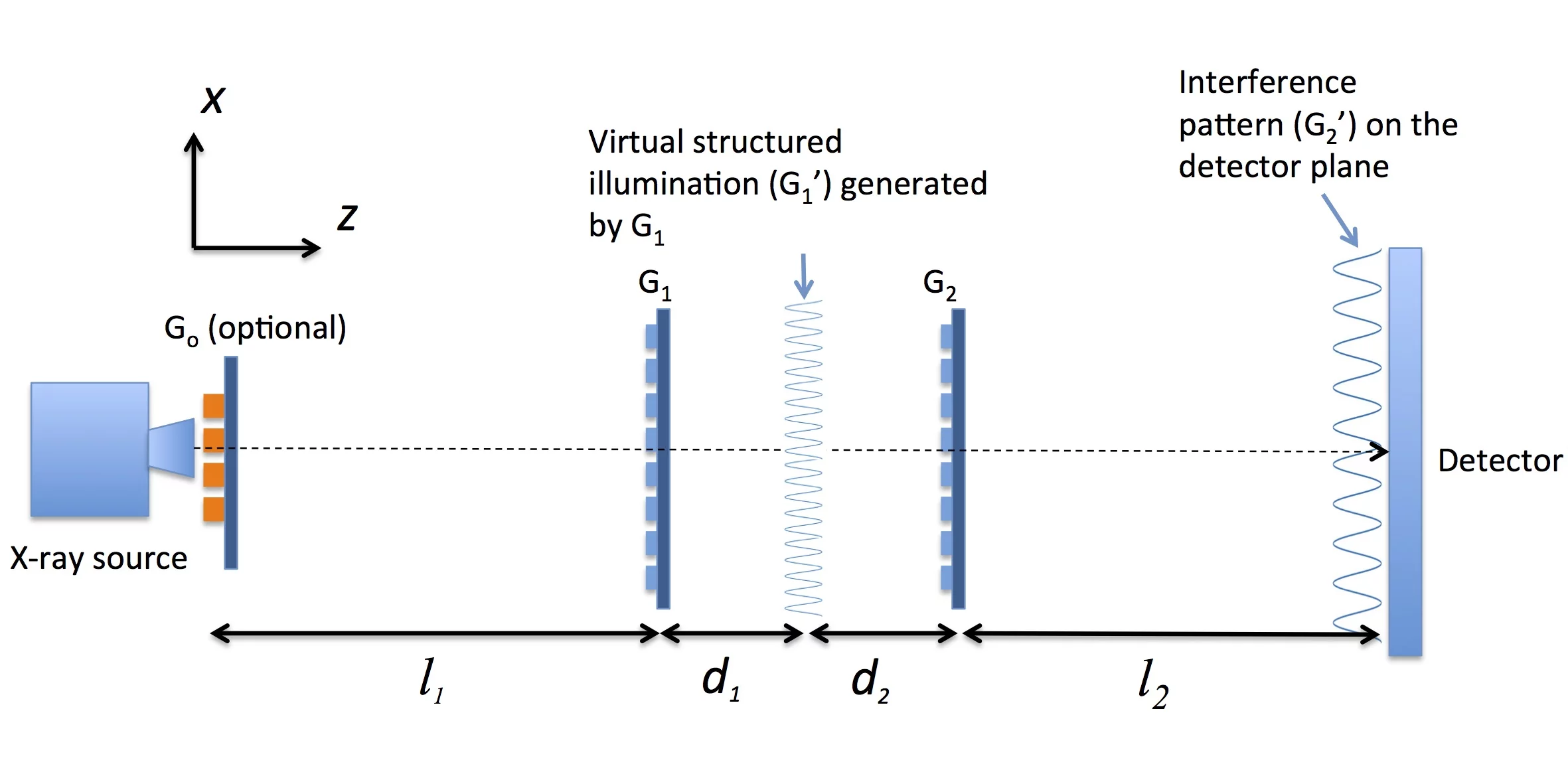
Dual Phase Grating Interferometer

Sketch of experimental setup.
This project explores the possibility of achieving applicable grating interferometer (GI) for table-top applications using only phase-shift gratings. The motivation is to remove the necessity of absorption gratings in conventional GI, which reduces the dose and flux efficient, and in the mean time bypass the fabrication difficulties for manufacturing high quality absorption gratings. The proposed interferometer consists of two phase gratings of small pitches that are placed close to each other. The whole system can be considered as two interferometers in cascade. This configuration generates a large fringe than can be resolved directly without the need of an analyser grating. Moreover, the proposed method provides a practical and convenient way of changing the dark-field sensitivity compared to conventional GI. By simply tuning the integrating distance in the millimeter range can sense structural information of the sample in different length scale.

Publications:

  1. Kagias. M, Wang Z, Jefimovs K, Stampanoni M,. APPLIED PHYSICS LETTERS 110, 014105 (2017)

Funding agencies: ERC Grant ERC-2012-StG 310005-PhaseX

Contact: Dr. Zhentian Wang, zhentian.wang@psi.ch, WBBA/212, +41-56-310-5819

Omnidirectional Dark-Field Imaging with Circular Unit Cell Gratings
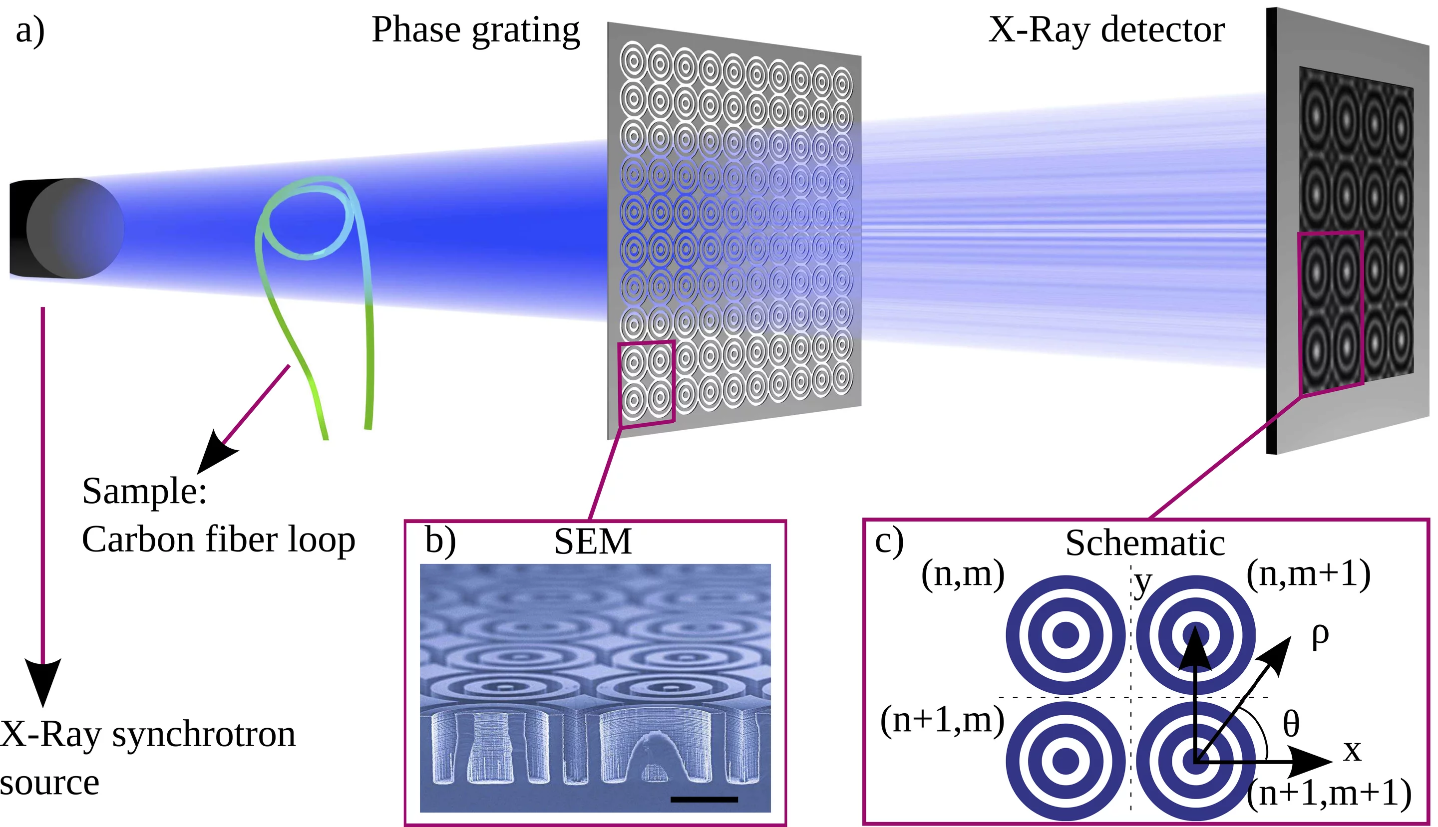
Sketch of experimental setup.
The dark-field signal in X-ray grating interferometry is highly directional, specifically for anisotropic samples the signal intensity depends on the angle between the grating lines and the sample. In this project a novel interferometric design is developed that allows 2D omnidirectional dark-field sensitivity. This is achieved by using fine pitch circular gratings that are repeated to cover the full field of view. The interference fringe generated by the dedicated grating is acquired and resolved with sufficient resolution, and then analysed. The gratings can be arranged in different configurations to optimize between the sensitivity and the final image resolution. The method has been validated [1] and benchmarked [2] at the TOMCAT beamline. On-going developments include the optimization of the analysis framework to increase directional sensitivity, translation of the imaging technique to a table-top setup, and ultimately the utilization of the method in tensor tomography experiments.

Publications:

  1. 2D-Omnidirectional Hard-X-Ray Scattering Sensitivity in a Single Shot Kagias. M, Wang Z, Villanueva-Perez P, Jefimovs K, Stampanoni M PHYSICAL REVIEW LETTERS 116, (2016). DOI: 10.1103/PhysRevLett.116.093902
  1. Circular Unit Cell Gratings for X-ray Dark-Field Imaging Kagias. M, Pandeshwar A.,Wang Z, Villanueva-Perez P, Jefimovs K, Stampanoni M JOURNAL OF PHYSICS: CONFERENCE SERIES submitted

Funding agencies: ERC Grant ERC-2012-StG 310005-PhaseX

Contact: Dr. Zhentian Wang, zhentian.wang@psi.ch, WBBA/212, +41-56-310-5819

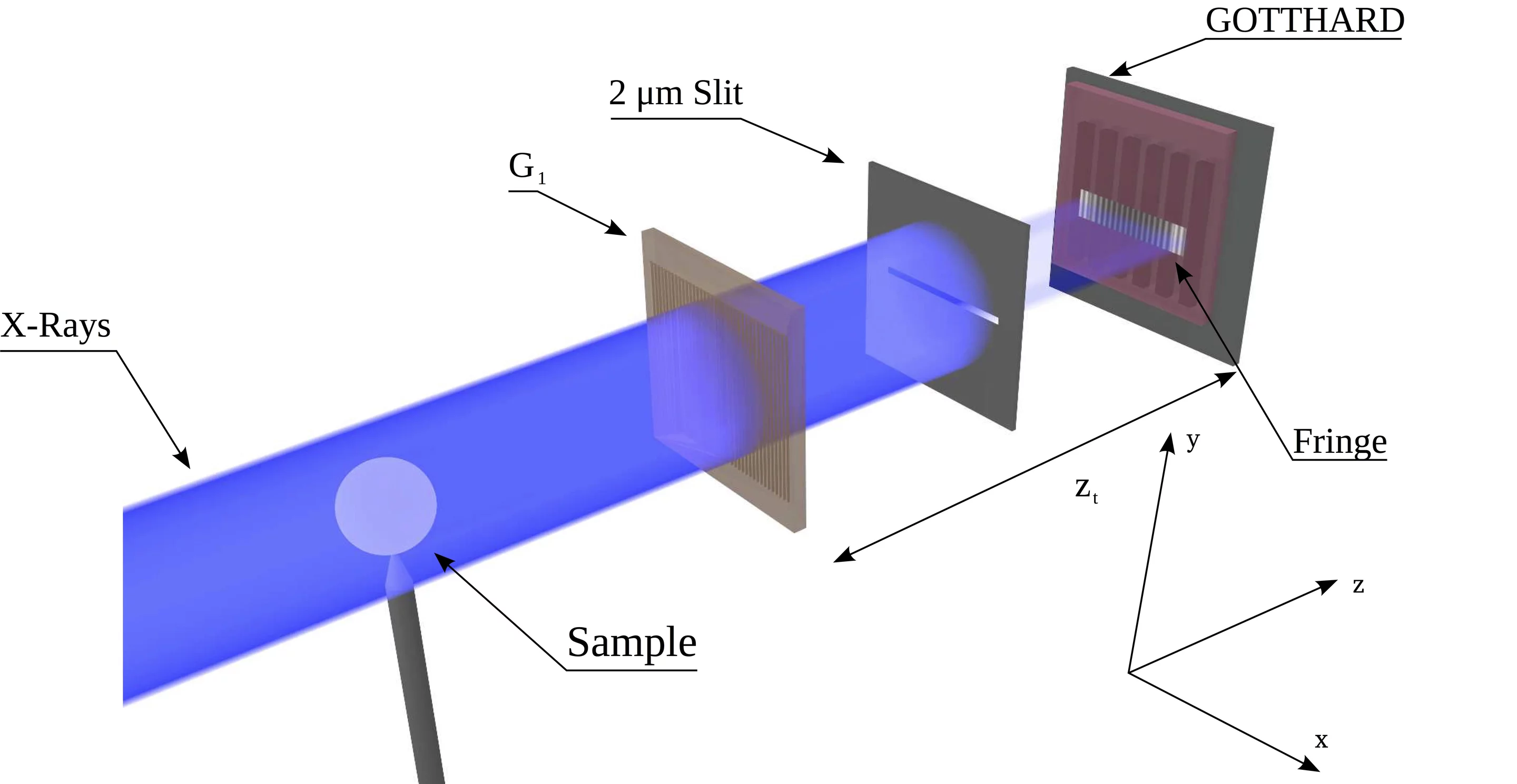
Single Shot Differential Phase Contrast Imaging with Single Photon Sensitive Detectors

Sketch of experimental setup.
This project focuses on the development of single shot grating based differential phase contrast imaging methods that do not require an analyser (absorption) grating. The removal of the analyser grating (known as G2) is favourable from both fabrication and photon utilization point of views. The proposed ‘G2-less’ imaging is achieved by utilizing a pixel interpolation method that allows resolution enhancement, which in turn is used to record interference fringe with very fine pitch directly. The resolution enhancement is achieved by exploiting the charge sharing that takes place in direct conversion detectors with small pixel sizes (25 micrometres). By exposing in a single photon sensitive regime the location of the incoming photon can be estimated with a precision higher than that dictated by the pixel size. The recorded fringe is then analysed by a custom phase retrieval algorithm based on the Hilbert transform. We have already managed to successfully demonstrate the applicability of the method with both strip (GOTTHARD) [1] and pixel (MOENCH) [2] detectors. Further developments, include the optimization of the imaging conditions and experiments with larger detector modules.

Publications:

  1. Micrometer-resolution imaging using MÖNCH: towards G 2 -less grating interferometry Cartier S, Kagias M, Bergamaschi A, Wang Z, Dinapoli R, Mozzanica A, Ramilli M, Schmitt B, Brückner M, Fröjdh E, Greiffenberg D, Mayilyan D, Mezza D, Redford S, Ruder C, Schädler L, Shi X, Thattil D, Tinti G, Zhang J, Stampanoni M JOURNAL OF SYNCHROTRON RADIATION 23, - (2016). DOI: 10.1107/S1600577516014788
  1. Single shot x-ray phase contrast imaging using a direct conversion microstrip detector with single photon sensitivity Kagias M, Cartier S, Wang Z, Bergamaschi A, Dinapoli R, Mozzanica A, Schmitt B, Stampanoni M APPLIED PHYSICS LETTERS 108, 234102 (2016). DOI: 10.1063/1.4948584

Collaboration:

  • SLS Detector Group, PSI, Switzerland

Funding agencies: ERC Grant ERC-2012-StG 310005-PhaseX

Contact: Dr. Zhentian Wang, zhentian.wang@psi.ch, WBBA/212, +41-56-310-5819

Differential phase contrast for X-ray tubes above 100 kVp

A 1D edge-on grating interferometer setup at PSI East.
Differential phase contrast and dark-field X-ray imaging have been developed over the last fifteen years. The applications have been extended from synchrotron sources to table-top systems with a Talbot-Lau geometry. Challenges in the fabrication of the optical components limited the deployment to sources typically used in mammography and cartilage screening, with an acceleration voltage below 40 kV. Applying these techniques to general purpose medical investigations or material analysis requires a re-evaluation of the performance and feasibility of grating interferometers on table-top sources, including a quantitative analysis of the response of the differential phase and dark-field signals related to the electron density and microstructural features of the sample.

Publications:

  1. X-ray phase-contrast imaging at 100 keV on a conventional source, Thüring T, Abis M, Wang Z, David C, Stampanoni M, Scientific Reports, 10.1038/srep05198, 2014.
  2. A generalized quantitative interpretation of dark-field contrast for highly concentrated microsphere suspensions Gkoumas S, Villanueva-Perez P, Wang Z, Romano L, Abis M, Stampanoni M, Scientific Reports, 10.1038/srep35259, 2016

Funding agencies: ERC Grant ERC-2012-StG 310005-PhaseX

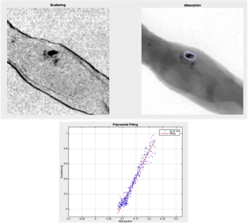
Ex-vivo study of suspicious microcalcifications in breast tissue biopsies

Absorption and scattering signals of microcalcification in breast biopsy tissue.
Breast cancer is the second most frequently diagnosed cancer in the world and it is the first leading cause of cancer-related deaths in women in less developed regions. The identification of (clustered) microcalcifications plays an important role in early detection of pre-malignant and malignant lesions. Microcalcifications comprise tiny calcium deposits possibly located in areas of accelerated cell turnover, suggestive of precancerous changes or early invasive breast cancer. The hypothesis of this project is that the chemical composition of microcalcifications as well as their internal microstructure are associated with the microenvironment in which the microcalcifications are formed. By probing these information, a descriptor could be established with the goal to improve breast cancer diagnosis. The project aims to understand the structural features of microcalcifications using X-ray small-angle scattering (SAXS) and ptychography, and eventually investigate the potential of using grating interferometer to classify ex-vivo microcalcifications using their absorption and dark-field (scattering) signals.

Publications:

[1] The first analysis and clinical evaluation of native breast tissue using differential phase-contrast mammography Stampanoni M, Wang Z, Thuering T, et al,, Invest. Radiol. 46(12):801, 2011

[2] Non-invasive classification of microcalcifications with phase-contrast X-ray mammography Wang Z, Hauser N, Singer G, et al., Nat. Commun. 5:3797m 2014

Funding agencies:

  • ERC Grant ERC-2012-StG 310005-PhaseX

Contact: Dr. Zhentian Wang, zhentian.wang@psi.ch, WBBA/212, +41-56-310-5819

Feasibility study of an X-ray phase contrast breast CT scanner

Sketch of a grating interferometer based breast CT scanner.
The aim of this project is to assess the feasibility of a grating interferometry (GI) based phase contrast breast CT system. While GI is an established tool for phase contrast X-ray CT imaging and is used for a wide range of applications, its transfer to clinically relevant systems in not obvious. Such a scanner needs to comply with strict dose requirements, while retaining a high level of sensitivity and accuracy. This requires operating the interferometer at higher X-ray energies, which poses challenges to all hardware components, in particular to high energy gratings and detectors. Additionally, the physical model behind high energy GI is not yet fully understood, further complicating any performance assessments. Implementing a grating interferometer on a fast rotating gantry demands high stability and consequently fast and robust signal retrieval methods. Thus, a general framework has to be developed that considers all mechanical, physical and hardware based limitations. This includes a performance analysis of signal retrieval methods and a comparison of absorption and phase contrast based imaging, as well as an analytical and simulation based understanding of the image formation process and dose deposition. From the hardware point of view the feasibility of a highly stable CT scanner is assessed, as well as the resulting requirements on and capabilities of dedicated hardware components.

Funding agencies:


  • ERC-2012-StG 310005-PhaseX

Contact: Dr. Zhentian Wang, zhentian.wang@psi.ch, WBBA/212, +41-56-310-5819

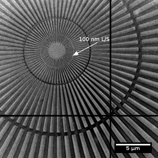
Fabrication of gratings for phase contrast X-ray imaging

Examples of gratings

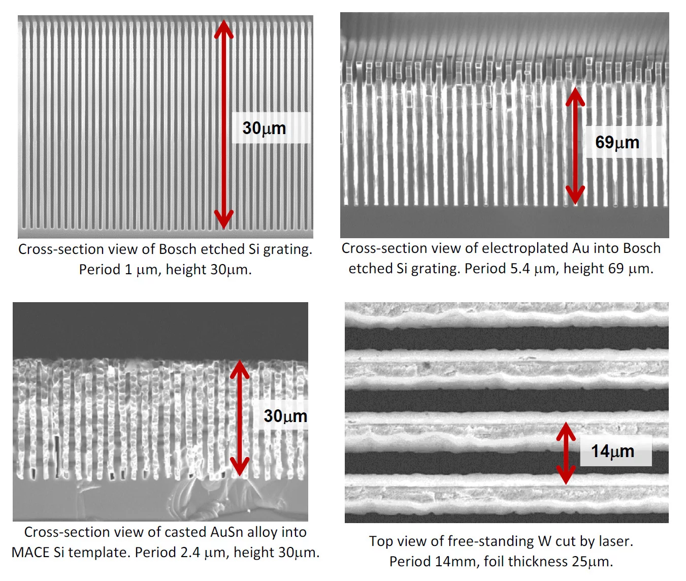
Grating interferometry is proved to be one of the most promising techniques for phase contrast X-ray imaging. Typical grating interferometer consists of a phase shifting grating (G1), analyzing grating (G2) and an optional absorbing source grating (G0). Usually, required grating period is in a range of few microns. The height of the lines of G1 grating should provide a certain phase shift, while the height of the grating lines of G2 should be sufficient to suppress radiation of defined energy. In both cases, structures with heights of tens (or even hundreds) of micrometers are required. However, the realization of structures with so high aspect ratios yet having sufficient quality over the large area is demanding. We develop fabrication procedures which enable such gratings. We produce G1 gratings in Si by reactive ion etching using Bosch technique [1] or by metal assisted chemical etching [2]. The absorbing G0 and G2 gratings are produced by filling Si templates with metal utilizing electroplating, metal casting [3] or atomic layer deposition [4]. Larger structures can alternatively be produced by laser cutting in W foils.

Publications:

  1. K. Jefimovs et al., “High aspect ratio silicon structures by Displacement Talbot lithography and Bosch etching“ Proc. SPIE (submitted 2017).
  2. L. Romano et al., Self-assembly nanostructured gold for high aspect ratio silicon microstructures by metal assisted chemical etching, RSC Advances 6 (2016) 16025-16029.
  3. L. Romano et al., “High aspect ratio metal microcasting by hot embossing for X-ray optics fabrication“ Microelectron. Eng. 17 (2017) 6-10.
  4. K. Jefimovs et al., “Zone-Doubling Technique to Produce Ultrahigh-Resolution X-Ray Optics” Phys. Rev. Lett. 99 (2007) 264801.

Collaboration:

  • Dr. Rolf Brönnimann, EMPA Dübendorf

Contacts: Dr. Konstantins Jefimovs, konstantins.jefimovs@psi.ch, WBBA/220, +41 56 310 3713 * Dr Lucia Romano, lucia.romano@psi.ch, WBBA/220, +41 56 310 5688