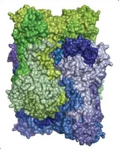
The small angle diffraction of hard x-rays provides statistical information on nanoscale ordering and structure. We develop experimental methods for time resolved measurement of structural changes induced by laser radiation, microfluidic mixing or temperature changes. Image on the left shows interlocked octameric rings of CS2 hydrolase (abstract in journal webpage).
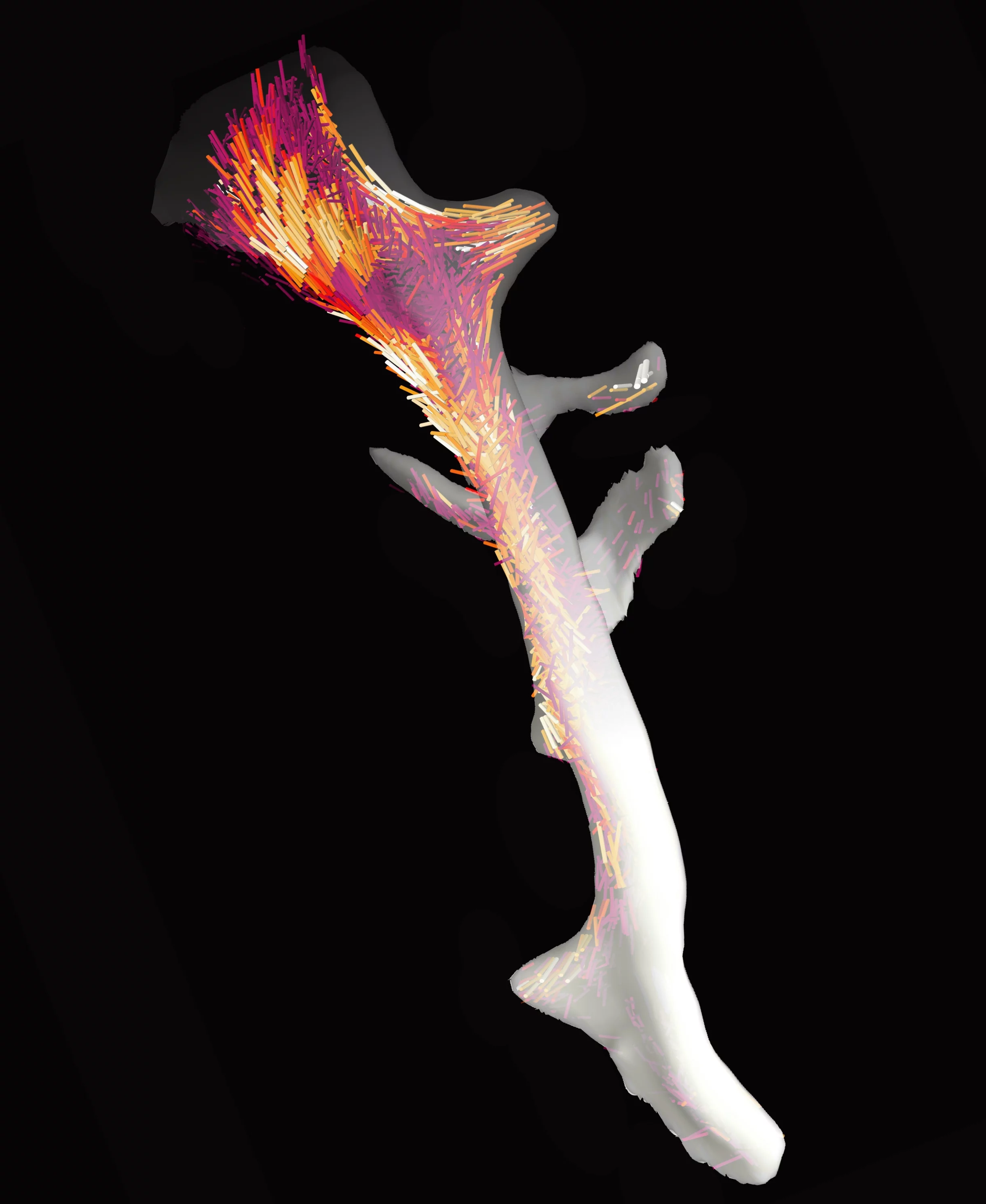
By scanning the sample relative to the x-ray beam we obtain spatially resolved SAXS datasets. For each point in the images a statistical description of ordering in the nanoscale is available. This technique can be further combined with sample rotations to probe ordering in 3D or to obtain measurements that are resolved in three-dimensions via tensor computed tomography (CT). In the image on left we show the result on a trabecular bone fragment of roughly two and a half millimetre long. The image shows a spatially resolved representation of the arrangement, orientation, and degree of orientation of nanoscale collagen fibrils (abstract in journal webpage).
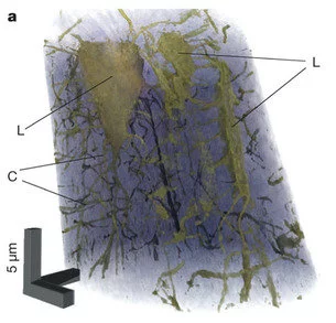
Ptychography - Scanning x-ray diffraction microscopy
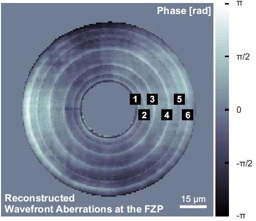
Using iterative algorithms as computational lenses coherent x-ray diffraction patterns are phased and render quantitative phase and amplitude images of the sample under study. We investigate and develop sample environments, measurement schemes, and reconstruction algorithms to further the development and application of this technique. Shown on the left is a 3D quantitative reconstruction of a small section of mouse femur bone, highlighting the osteocyte lacunae (L) and the connecting canaliculi network (C) (abstract in journal webpage).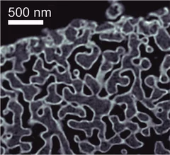
OMNY - tOMography Nano crYo stage
We work closely to the OMNY project in developing and testing a dedicated instrument for high-resolution scanning tomography based on differential interferometry. Two instruments exist, one in air and room temperature for added flexibility in the measurement environment, and a second one in vacuum and cryogenic temperatures to allow measurements on radiation sensitive materials such as soft tissue. This method achieves a stability of better than 10 nm between beam defining optics and sample. As an example we show on the left a tomogram of a coated nanoporous glass sample where three distinct gray levels are visible for air (black), glass (gray), and Ta2O5 (white). The projections were measured using ptychography and an isotropic 3D resolution of 16 nm was achieved (abstract in journal webpage).