Mapping the Nanoscale Architecture of Functional Materials
A new X-ray technique reveals the 3D orientation of ordered material structures at the nanoscale, allowing new insights into material functionality.
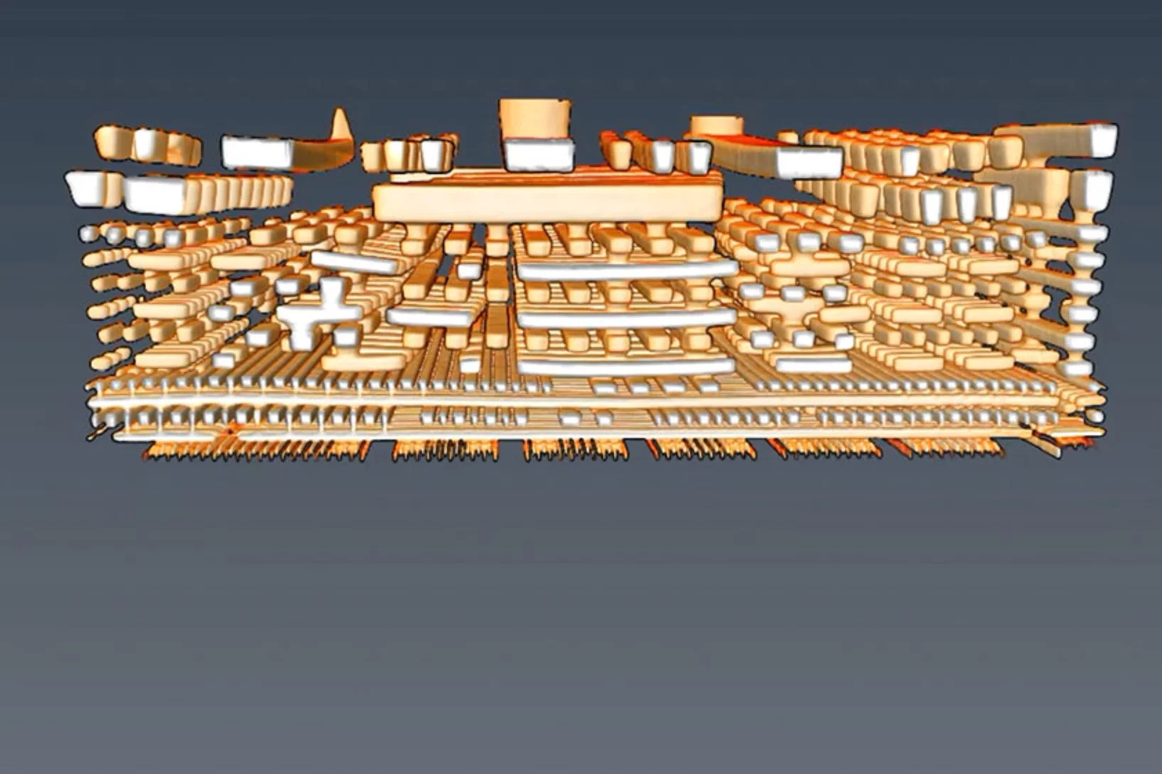
Neuer Röntgenweltrekord: Blick in einen Computerchip auf 4 Nanometer genau
Mit einer Rekordauflösung von 4 Nanometern gelang es Forschenden am PSI, die räumliche Struktur eines Computerchips mithilfe von Röntgenlicht abzubilden.
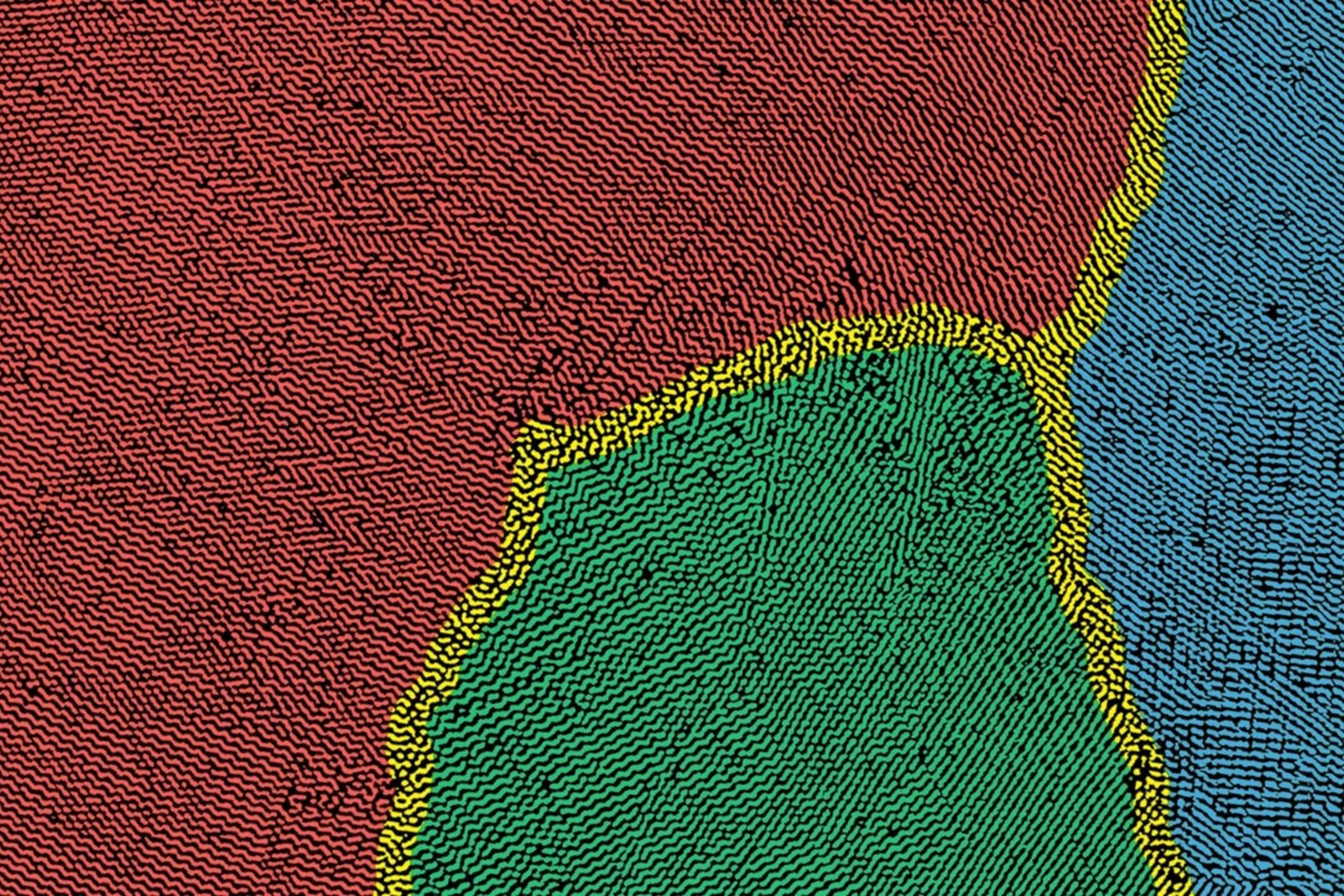
Nanoimaging Reveals Topological Textures in Nanoscale Crystalline Networks
X-ray nano-tomography reveals collective behavior in synthetic self-assembled nanostructures. The new method opens opportunities for the synthesis of photonic and plasmonic materials with improved long-range ordering.
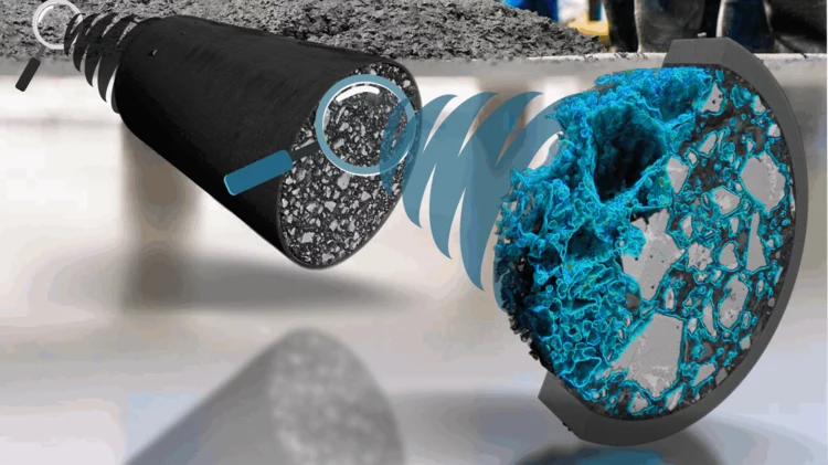
A deep look into hydration of cement
Researchers led by the University of Málaga show the Portland cement early age hydration with microscopic detail and high contrast between the components. This knowledge may contribute to more environmentally friendly manufacturing processes.
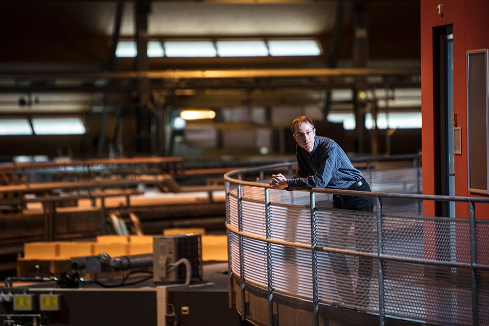
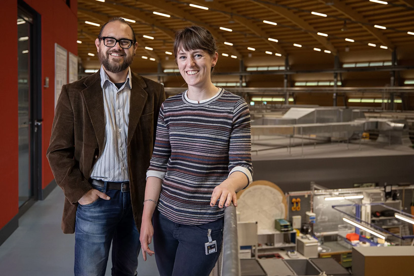
Manuel Guizar-Sicairos appointed as Associate Professor at EPF Lausanne and head of the Computational X-ray Imaging group at PSI
Dr. Manuel Guizar-Sicairos, currently Senior Scientist at PSI, was appointed as Associate Professor of Physics in EPF Lausanne and head of the Computational X-ray Imaging group in PSI.
Dr. Manuel Guizar-Sicairos elected as SPIE Fellow member
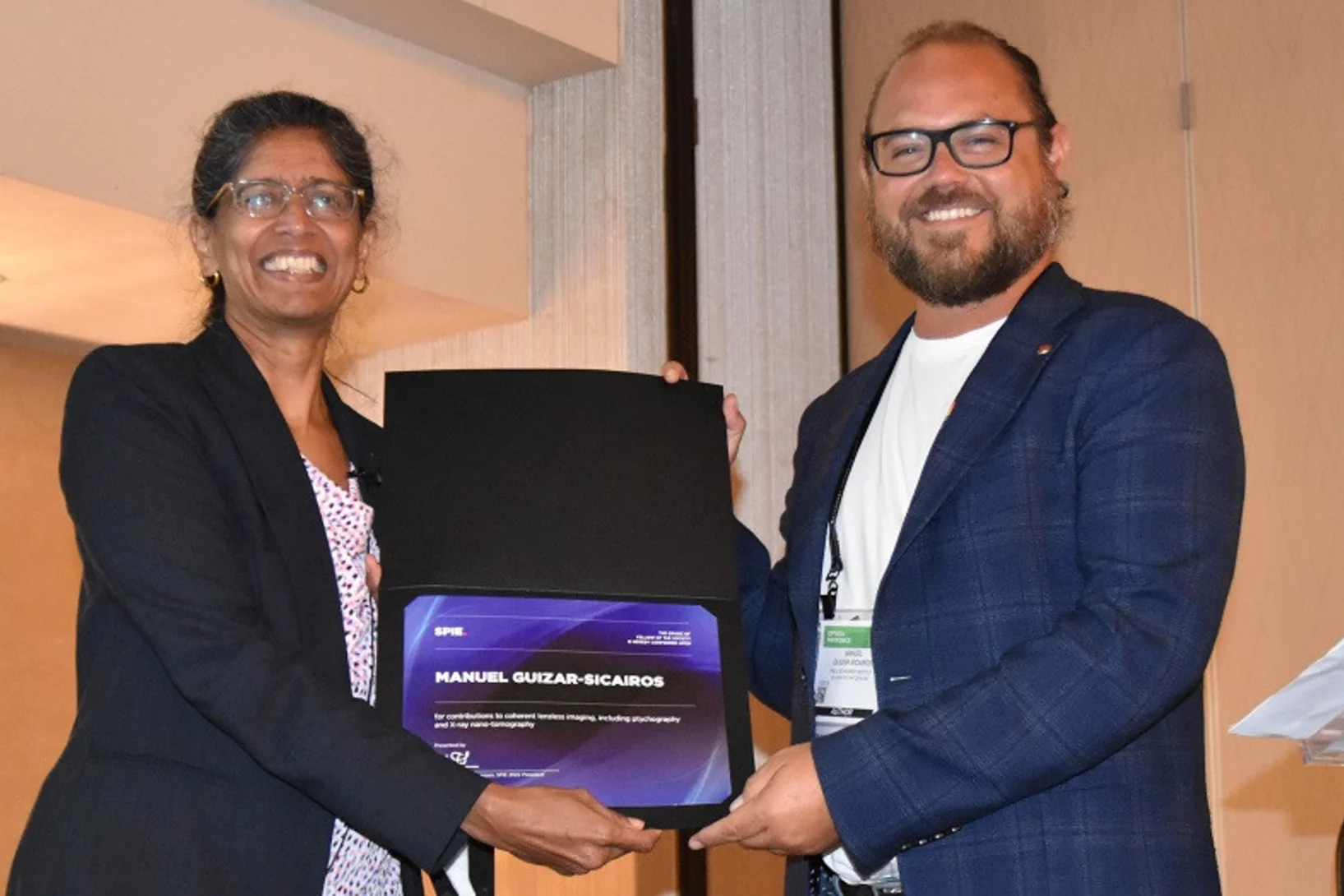
Dr. Manuel Guizar-Sicairos was elected as a 2022 SPIE Fellow Member for his contributions to coherent lensless imaging, including ptychography and X-ray nano-tomography. The distinction was awarded in the SPIE’s Optics & Photonics conference in San Diego, California.
Nanomaterial aus dem Mittelalter
Das Geheimnis des Zwischgolds wurde am PSI gelüftet.
Spiegelbildliche Moleküle leichter unterscheiden
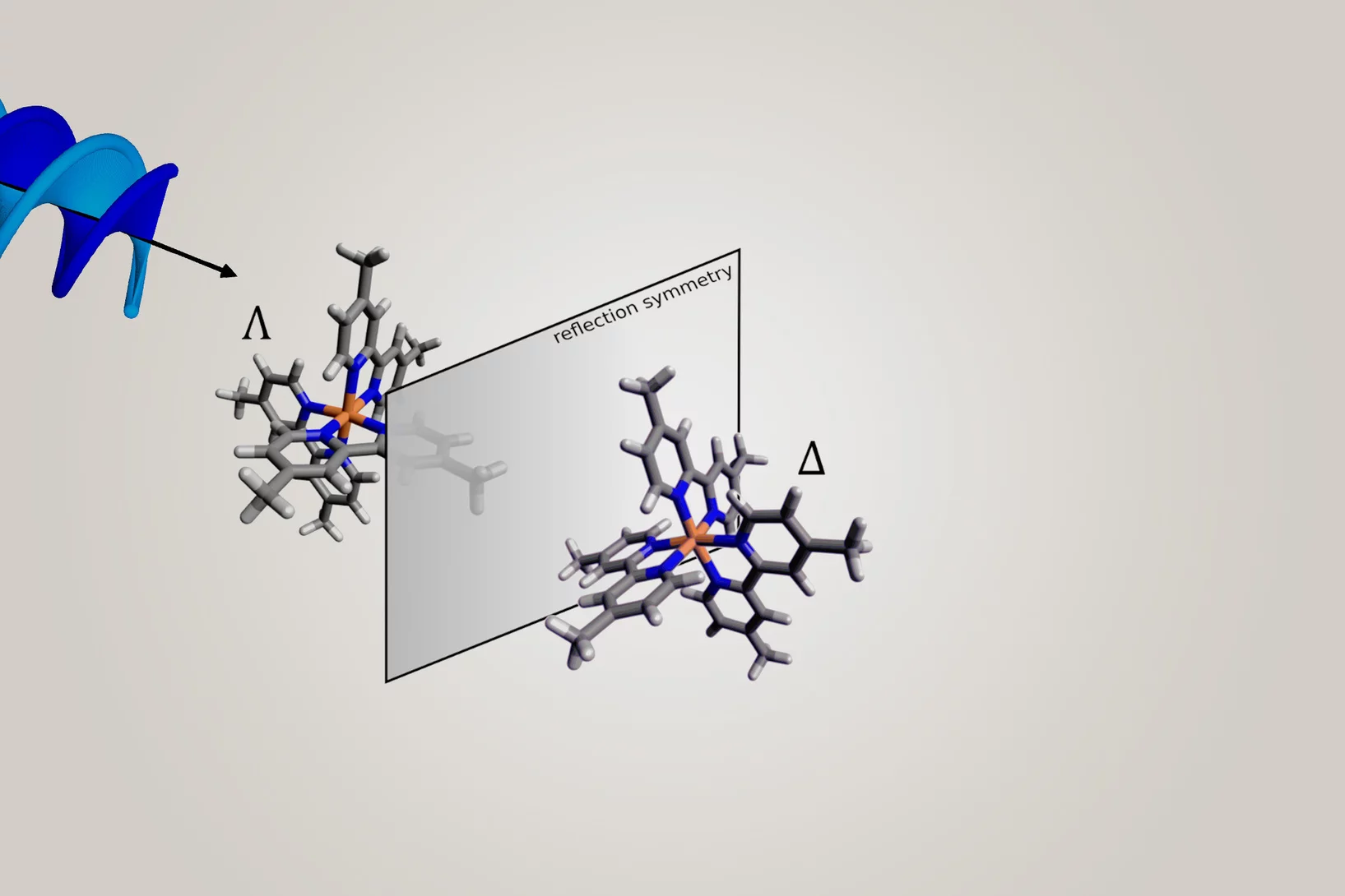
Mit schraubenförmigem Röntgenlicht lassen sich spiegelbildliche Substanzen – sogenannte Enantiomere – besser voneinander unterscheiden.
Neuartige Röntgenlinse erleichtert Blick in die Nanowelt
Erstmals gibt es nun eine achromatische Linse auch für Röntgenlicht – entwickelt am PSI.
Revealing invisible defects in fusion reactor armor
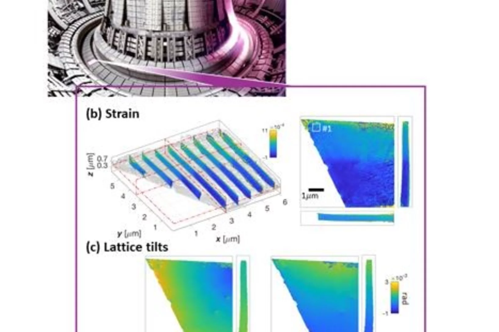
In an exciting collaboration, Nick Phillips, a PSI Fellow at the cSAXS beamline, reveals nanoscale lattice distortions created by invisible defects in fusion reactor armor. This work develops the current understanding of how the smallest, but most prevalent defects, generated during neutron irradiation behave. The novel Bragg ptychographic approach published in Nature Communications paves the way for fast, robust, 3D Bragg ptychography.
Ground-breaking technology development recognised
PSI researchers win the international Innovation Award on Synchrotron Radiation for 3D mapping of nanoscopic details in macroscopic specimens, such as bone.
Neurodegenerative disease studied by cryogenic X-ray nanotomography
Hard X-ray cryo-tomography scanning of retina from healthy and inherited blindness specimen paves the way for correlative analysis after imaging at the cSAXS beamline.
Dr. Manuel Guizar-Sicairos is awarded ICO prize
Dr. Manuel Guizar-Sicairos, beamline scientist at the cSAXS beamline, is the 2019 recipient of the International Commission for Optics (ICO) Prize. The distinction was awarded in the EOSAM conference in Rome.
Imaging strain with high resolution
Imaging strain in crystalline materials with high resolution can be a challenging task. Researchers demonstrate an original use of X-ray ptychography for this purpose: ptychographic topography.
Wie Katalysatoren altern
Katalysatoren, die in der Industrie eingesetzt werden, verändern über die Jahre ihre Materialstruktur. Mit einer neuen Methode haben PSI-Forschende dies nun auf der Nano-Skala untersucht.
Understanding Why Solid-State Batteries Fail
Researchers from the University of Oxford, the Diamond Light Source and the Paul Scherrer Institut have generated strong evidence supporting one of two competing theories regarding the mechanism by which lithium metal dendrites grow through ceramic electrolytes. A process leading to short circuit at high rates of charge. The X-ray phase-contrast imaging capabilities of the TOMCAT beamline of the Swiss light source allowed researchers to visualize and characterize the growth of cracks and dendrites deep within an operating solid-state battery. The results were published in Nature Materials on April 22, 2021.
Quantifying oriented myelin in mouse and human brain
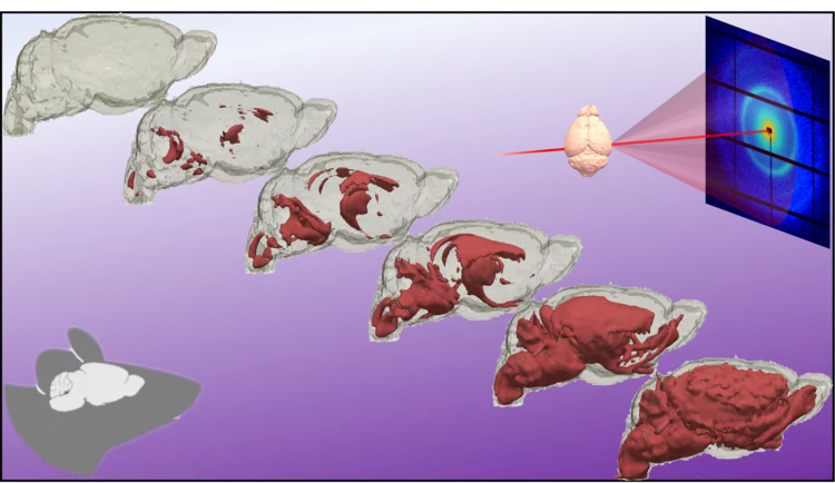
Myelin 'insulates' our neurons enabling fast signal transduction in our brain. Myelin levels, integrity, and neuron orientations are important determinants of brain development and disease. Small-angle X-ray scattering tensor tomography (SAXS-TT) is a promising technique for non-destructive, stain-free imaging of brain samples, enabling quantitative studies of myelination and neuron orientations, i.e. of nano-scale properties imaged over centimeter-sized samples.
Magnetic vortices come full circle
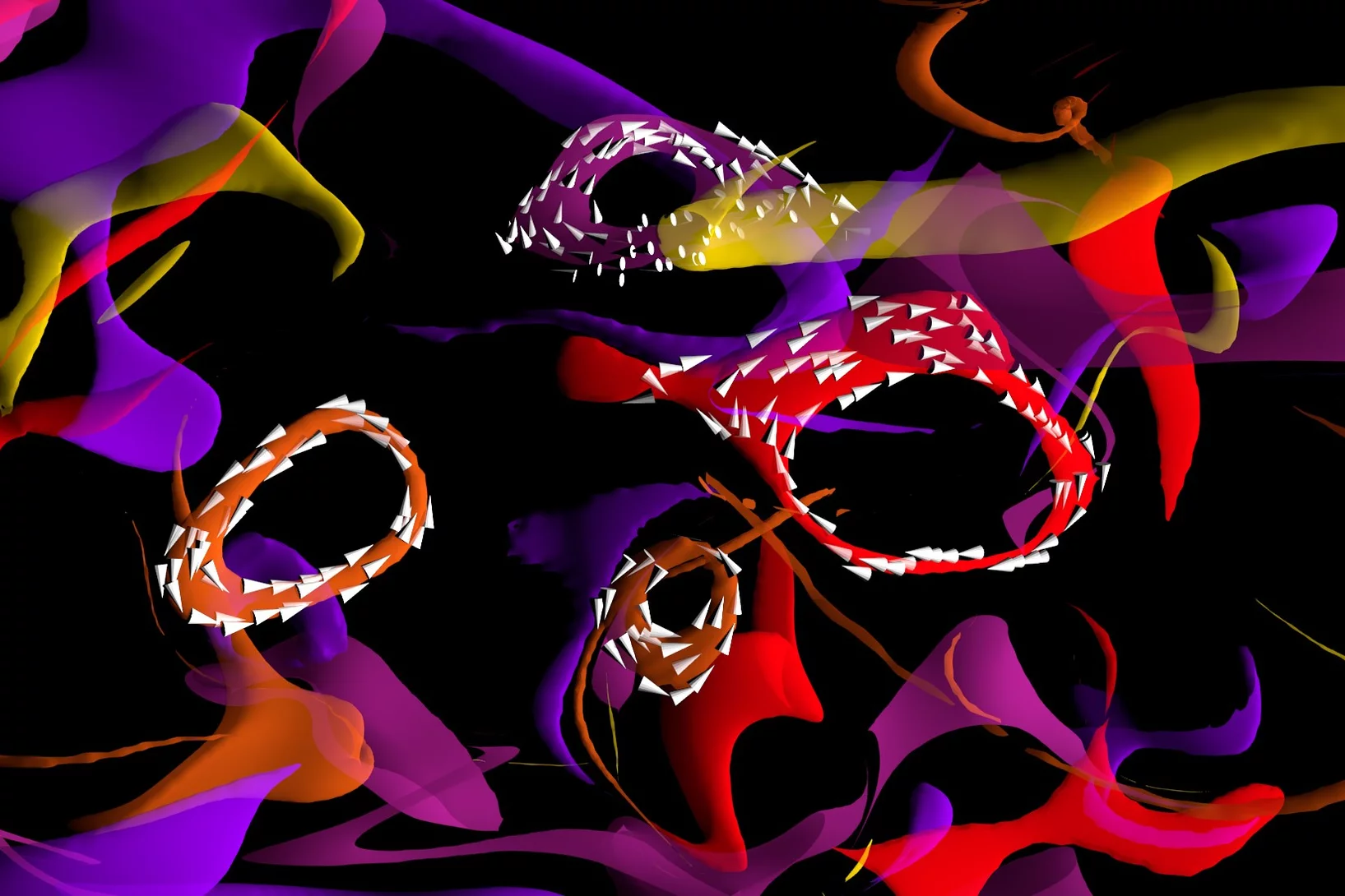
The first experimental observation of three-dimensional magnetic ‘vortex rings’ provides fundamental insight into intricate nanoscale structures inside bulk magnets, and offers fresh perspectives for magnetic devices.
Dr. Manuel Guizar-Sicairos elected as Fellow member of The Optical Society (OSA)
Dr. Manuel Guizar-Sicairos, beamline scientist at the cSAXS beamline, was elected as a Fellow Member of The Optical Society (OSA) for seminal contributions to methods and applications of coherent lensless imaging, ptychography, x-ray nanotomography, and new modalities of x-ray microscopy.
Nanowelten in 3-D
Tomogramme aus dem Inneren von Fossilien, Hirnzellen oder Computerchips liefern neue Erkenntnisse über feinste Strukturen. Die 3-D-Bilder gelingen mithilfe der Röntgenstrahlen der Synchrotron Lichtquelle Schweiz SLS dank eigens entwickelter Detektoren und raffinierter Computeralgorithmen.
Kurzfilm eines magnetischen Nanowirbels
Mit einer neu entwickelten Untersuchungsmethode konnten Forschende die magnetische Struktur im Inneren eines Materials mit Nanometer-Auflösung abbilden. Ihnen gelang ein kurzer «Film» aus sieben Bildern, der erstmalig in 3-D zeigt, wie sich winzige Wirbel der Magnetisierung tief im Inneren eines Materials verändern.
Soft X-ray Laminography: 3D imaging with powerful contrast mechanisms
3D imaging using synchrotron radiation is a widely used tool that allows access to the inner structure of complex objects. An international and interdisciplinary consortium of scientists from the Swiss Light Source (PolLux and cSAXs), the Friedrich-Alexander-Universität Erlangen-Nürnberg, and the University of Cambridge developed the new 3D imaging technique of Soft X-ray Laminography (SoXL). SoXL allows for the investigation of thin and extended samples while taking advantage of the characteristic absorption contrast mechanisms in the soft X-ray range, providing 3D information with nm spatial resolution.
3D imaging for planar samples with zooming
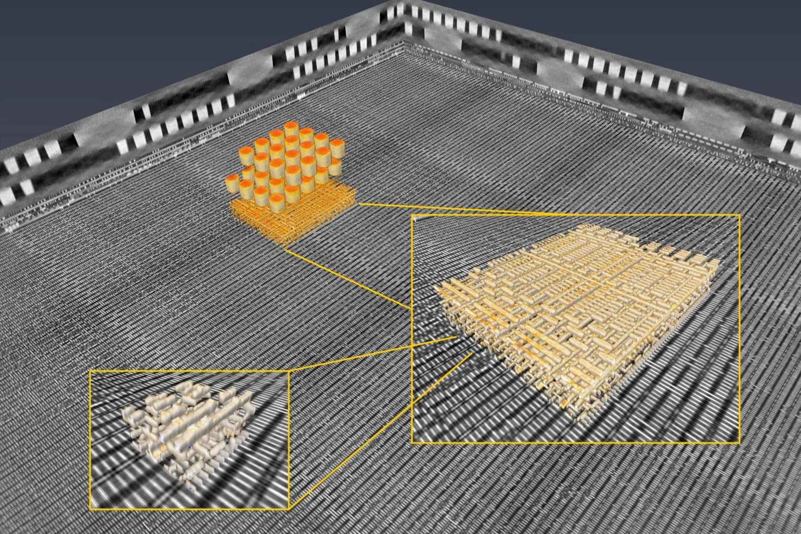
Researchers of the Paul Scherrer Institut have previously generated 3-D images of a commercially available computer chip. This was achieved using a high-resolution tomography method. Now they extended their imaging approach to a so-called laminography geometry to remove the requirement of preparing isolated samples, also enabling imaging at various magnification. For ptychographic X-ray laminography (PyXL) a new instrument was developed and built, and new data reconstruction algorithms were implemented to align the projections and reconstruct a 3D dataset. The new capabilities were demonstrated by imaging a 16 nm FinFET integrated circuit at 18.9 nm 3D resolution at the Swiss Light Source. The results are reported in the latest edition of the journal Nature Electronics. The imaging technique is not limited to integrated circuits, but can be used for high-resolution 3D imaging of flat extended samples. Thus the researchers start now to exploit other areas of science ranging from biology to magnetism.
Glycation of collagen in decellularized pericardium tissue: pilot study
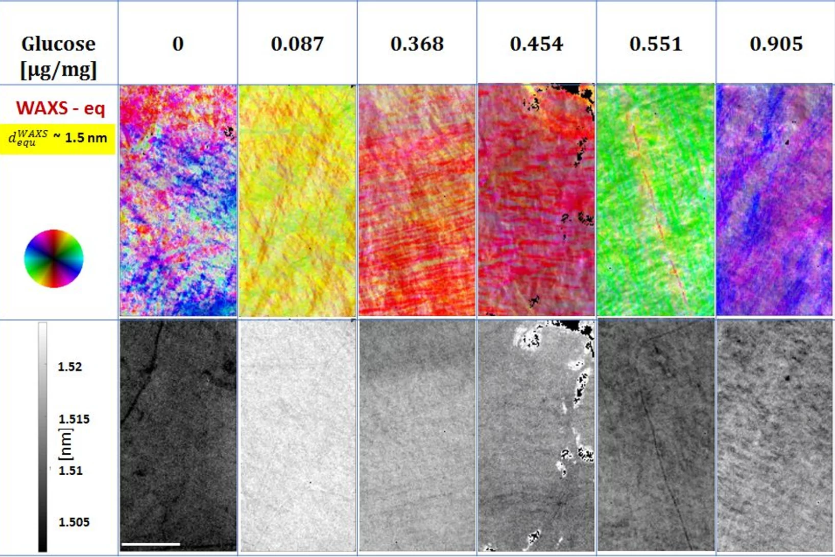
Aging populations and diabetics suffer from the effects of the glycation of collagen, the non-enzymatic formation of glucose bridges. While the secondary effects have been studied intensely, comparatively little is known on the direct effect of glycation on the structure of collagen. It has been demonstrated in this study, that the direct impact of glycation can be determined with sub-atomic precision, and that a model system based on abundantly available connective tissue of farm animals can be used for such studies. This opens new avenues for inspecting the effects of diabetes mellitus on connective tissues and the influence of therapies on the resulting secondary disorders.
Inside Batteries
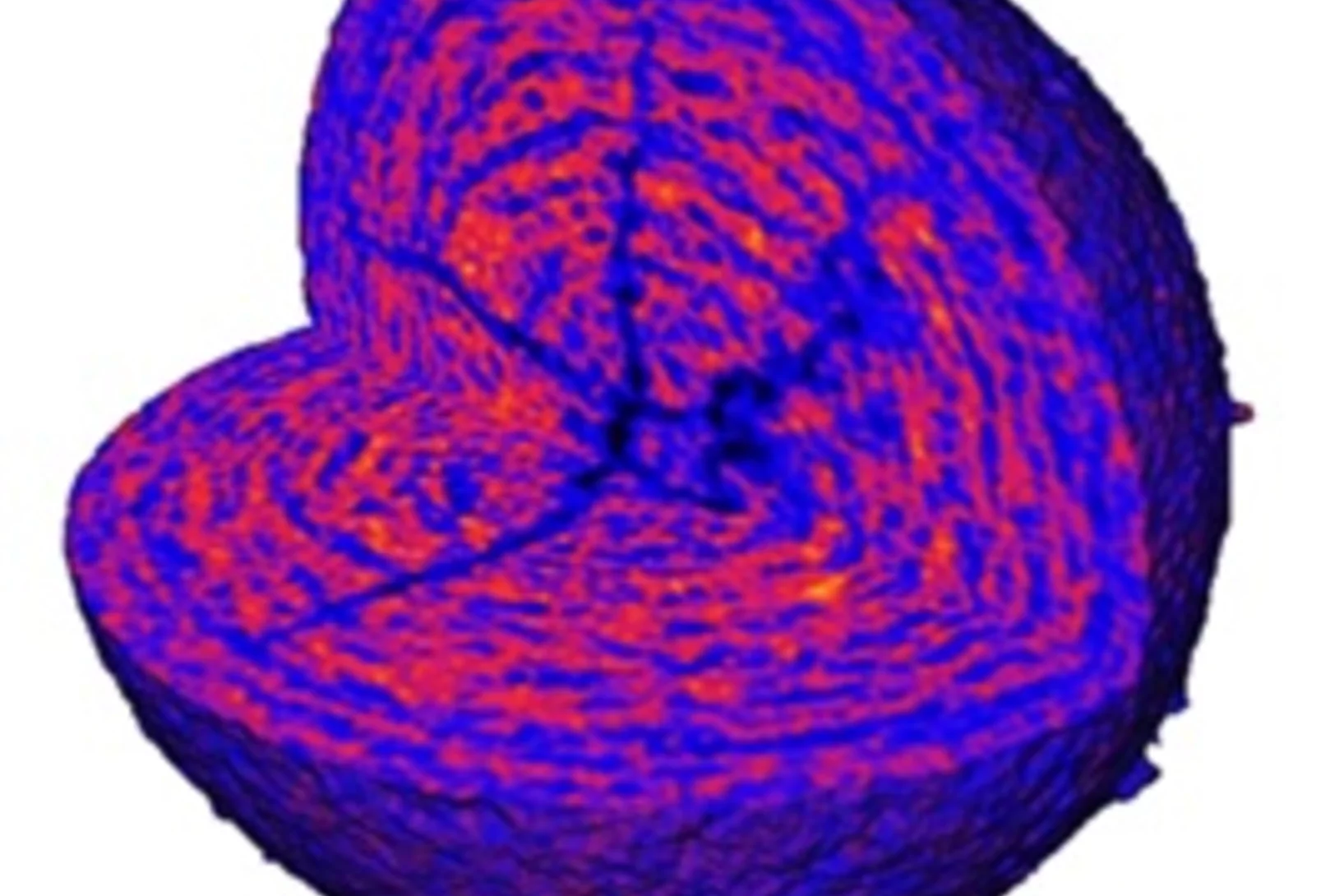
Lithium ion batteries (LIB) are essential in modern everyday life, with increasing interest in enhancing their performance and lifetime. Secondary particles of Li-rich cathode material were examined with correlated ptychographic X-ray tomography and diffraction microscopy at different stages of cycling to probe the aging mechanism.
Insights into a well-known disease in ageing populations: Abdominal and popliteal aneurysm
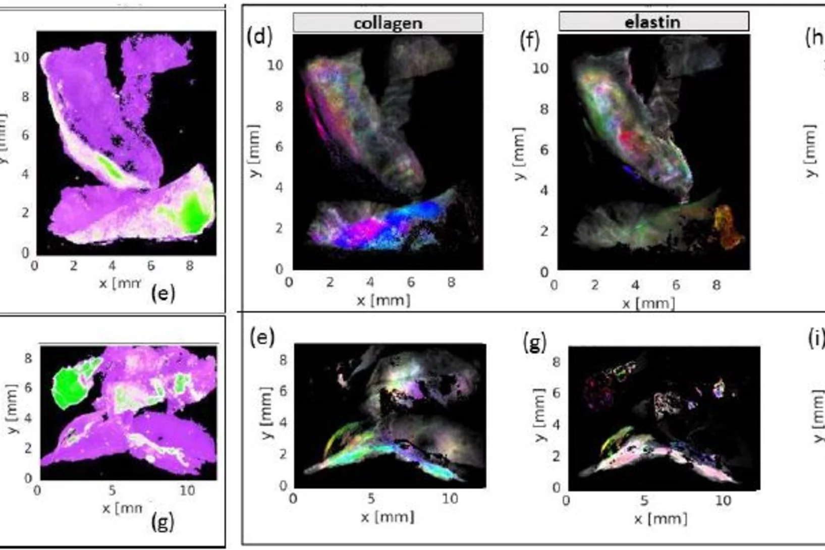
Abdominal aortic aneurysm, an enlargement of the abdominal aorta, may lead to rupture and thus acute health issues and death. Scanning X-ray imaging enabled new insights in the nano-structure of calcifications associated with abdonimal and popliteal aneurysm and allowed mapping the distribution of nano- and micro-calcifications as well as of collagen, elastin and myofilament as building blocks of connective tissue across samples from human donors.
Virtuelle Linse verbessert Röntgenmikroskopie
Eine von PSI-Forschenden neu entwickelte Methode macht Röntgenbilder von Materialien noch besser. Die Forschenden bewegten dafür eine optische Linse und nahmen dabei Einzelbilder auf. Mit Hilfe von Computeralgorithmen errechneten sie daraus ein Gesamtbild.
Helping chemists to understand degradation and stabilization of catalytic nanoporous gold structures
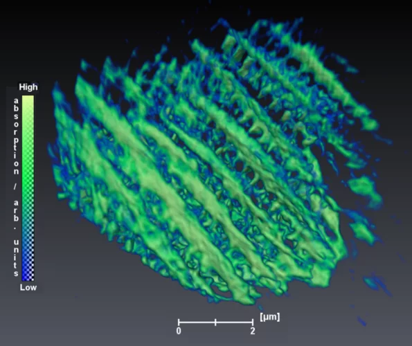
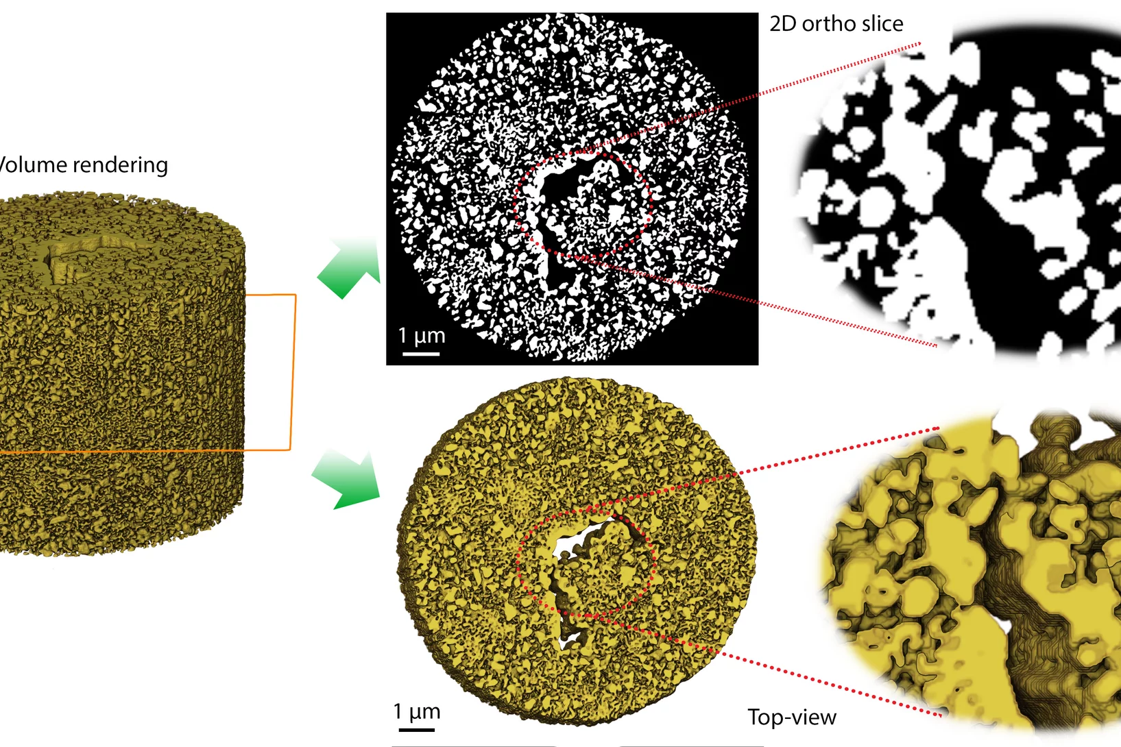
Catalytic materials are ubiquitously used in industrial processes to perform chemical reactions efficiently and in a sustainable manner. Nanoporous gold (npAu) is a monolithic sponge-like catalyst exhibiting a hierarchical structure with pores and connecting ligaments of typically 10 to 50 nm.
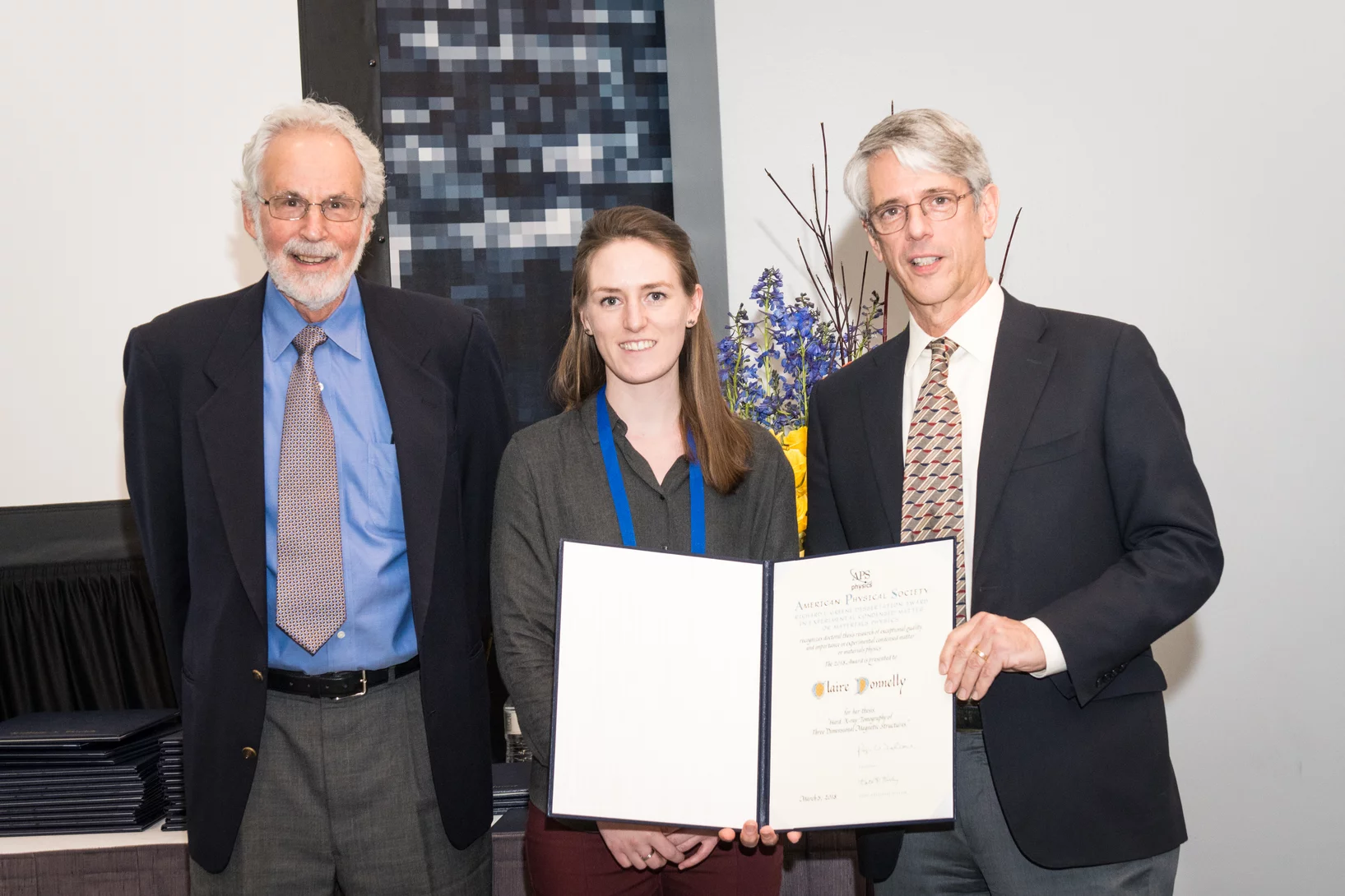
Claire Donnelly dissertation research awards
Claire Donnelly, Mesoscopic Systems (ETH Zurich - PSI), was awarded the COMSOL SPS Award in Computational Physics, the Werner Meyer-Ilse Memorial Award, the ETH Medal for an outstanding doctoral thesis, and the American Physical Society Richard L. Greene Dissertation Award.
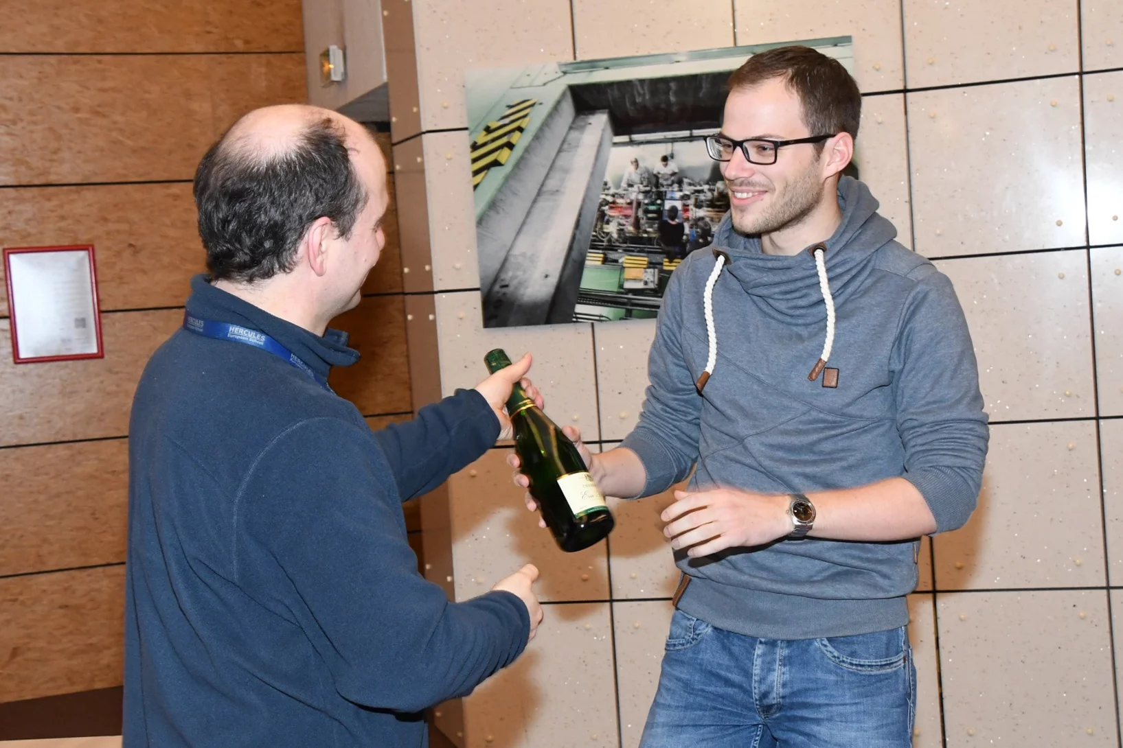
HERCULES School Poster Prize
Klaus Wakonig was awarded the best poster prize in the 2018 rendition of the HERCULES European School in Grenoble, France. Klaus is currently a PhD student at the CXS group at PSI, developing X-ray Fourier ptychography.

