Myelin “insulates” our neurons enabling fast signal transduction in our brain; myelin levels, integrity, and neuron orientations are important determinants of brain development and disease. However, myelin imaging methods used in clinics or research are non-specific or destructive.
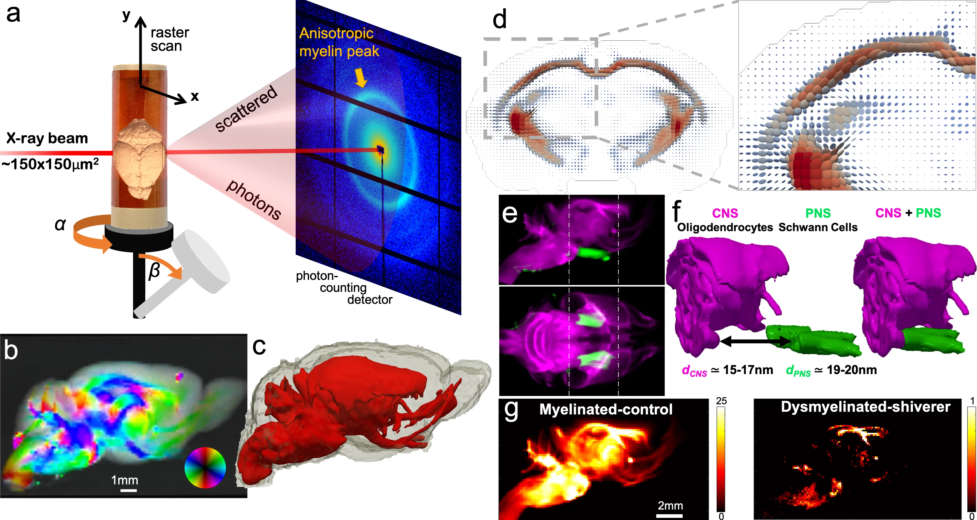
Using small-angle X-ray scattering tensor tomography (SAXS-TT), we exploited myelin’s ~17nm periodicity to non-invasively derive 3D myelin and neuron orientation maps in macroscopic tissue volumes (Figure). We demonstrated the method on a mouse brain (a-d), a mouse spinal cord, a human visual cortex and two human white matter specimens. We validated the readouts with 2D and 3D histology, and correlated the results with MRI contrasts.
Myelin’s ~3nm periodicity difference in the central and peripheral nervous system (CNS/PNS) enabled multiplexed imaging of the mouse brain and peripheral nerves (e,f). We also imaged control and diseased (dysmyelinated) mouse brains (g), highlighting SAXS-TT’s ability to tomographically image even minute amounts of myelin, and probe myelin structural integrity.
This non-destructive, stain-free imaging enables quantitative studies of myelination and neuron orientations within and across samples during development, aging, disease and treatment, and is applicable to other ordered biomolecules or nanostructures.
Contact
Dr. Marios Georgiadis
Stanford University
E-mail: mariosg@stanford.edu
Dr. Manuel Guizar-Sicairos
Paul Scherer Institut
E-mail: manuel.guizar-sicairos@psi.ch
Original Publication
Georgiadis, M., Schroeter, A., et al. Nanostructure-specific X-ray tomography reveals myelin levels, integrity and axon orientations in mouse and human nervous tissue. Nat. Commun. 12, 2941 (2021). doi: 10.1038/s41467-021-22719-7

