Mapping the Nanoscale Architecture of Functional Materials
A new X-ray technique reveals the 3D orientation of ordered material structures at the nanoscale, allowing new insights into material functionality.
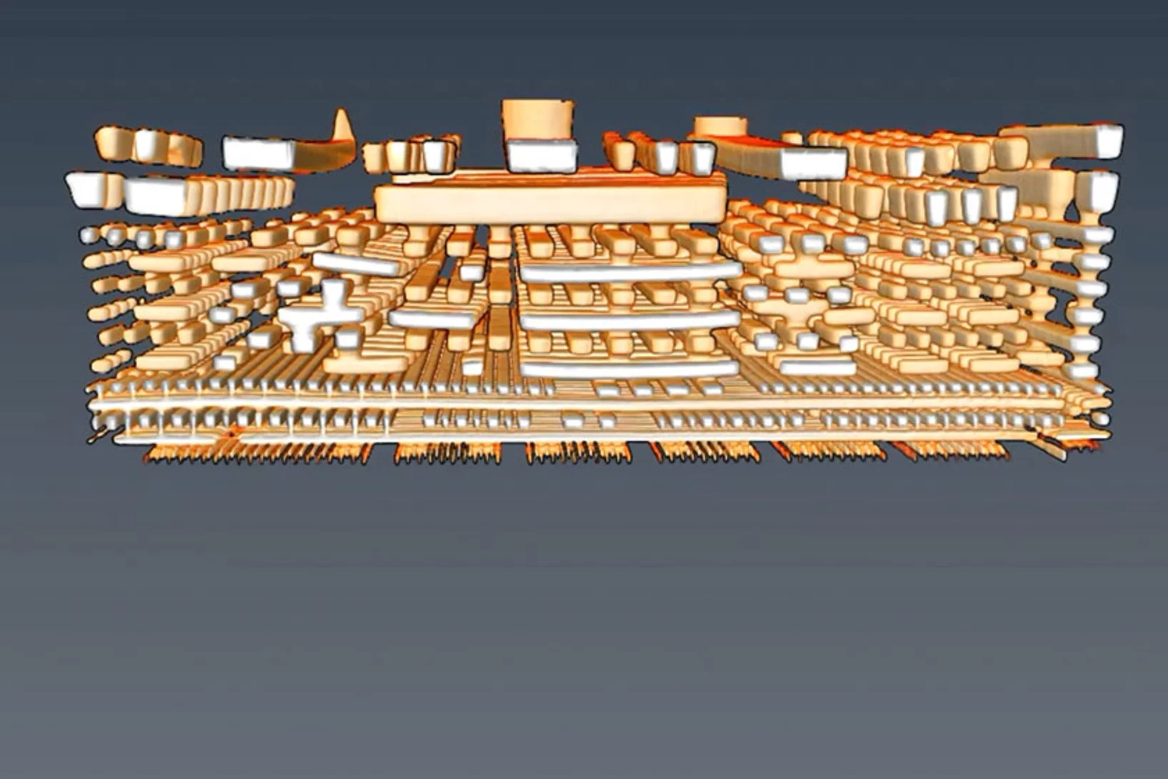
New X-ray world record: Looking inside a microchip with 4 nanometre precision
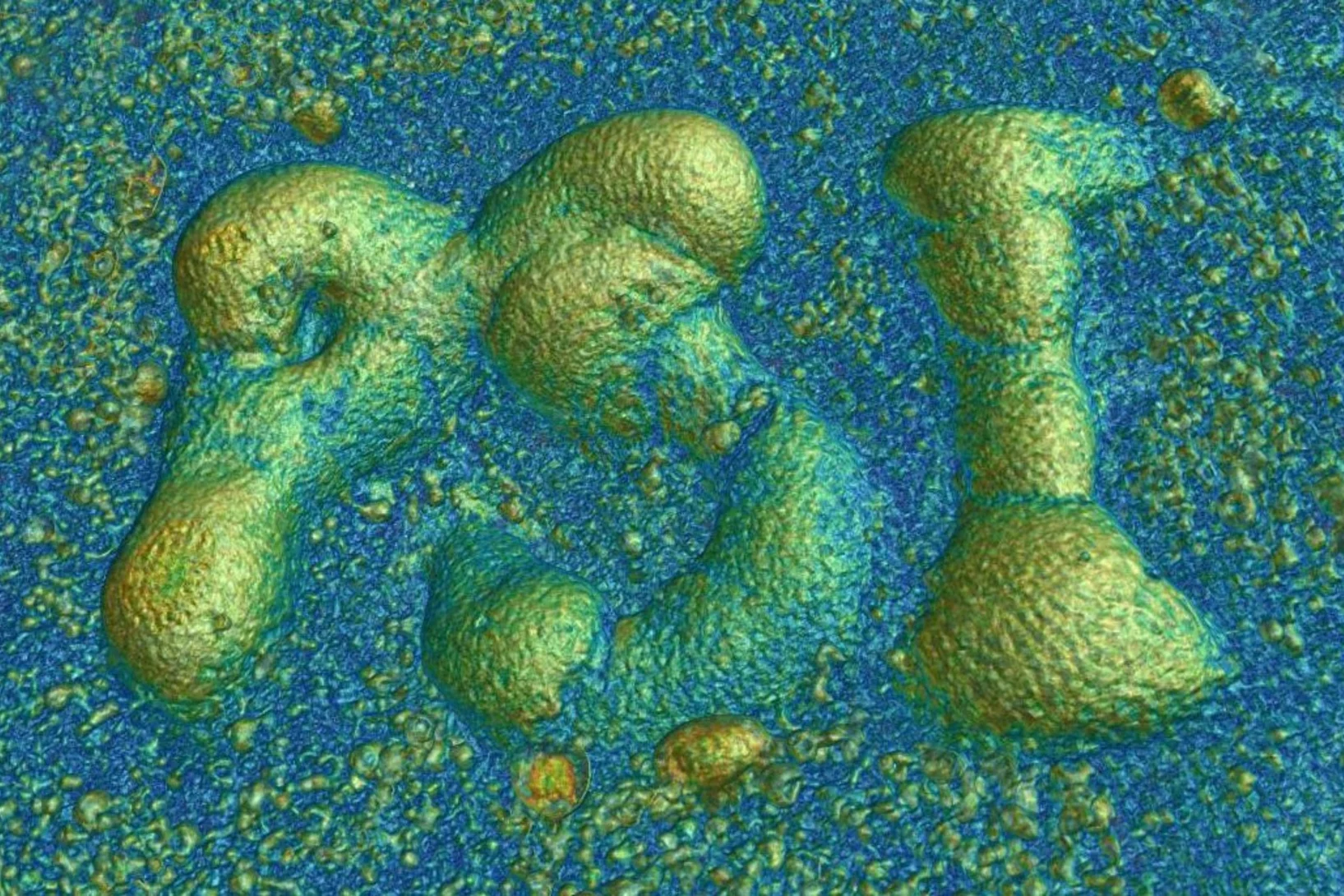
Researchers at PSI have succeeded in imaging the spatial structure of a computer chip with a record resolution of 4 nanometres using X-rays.
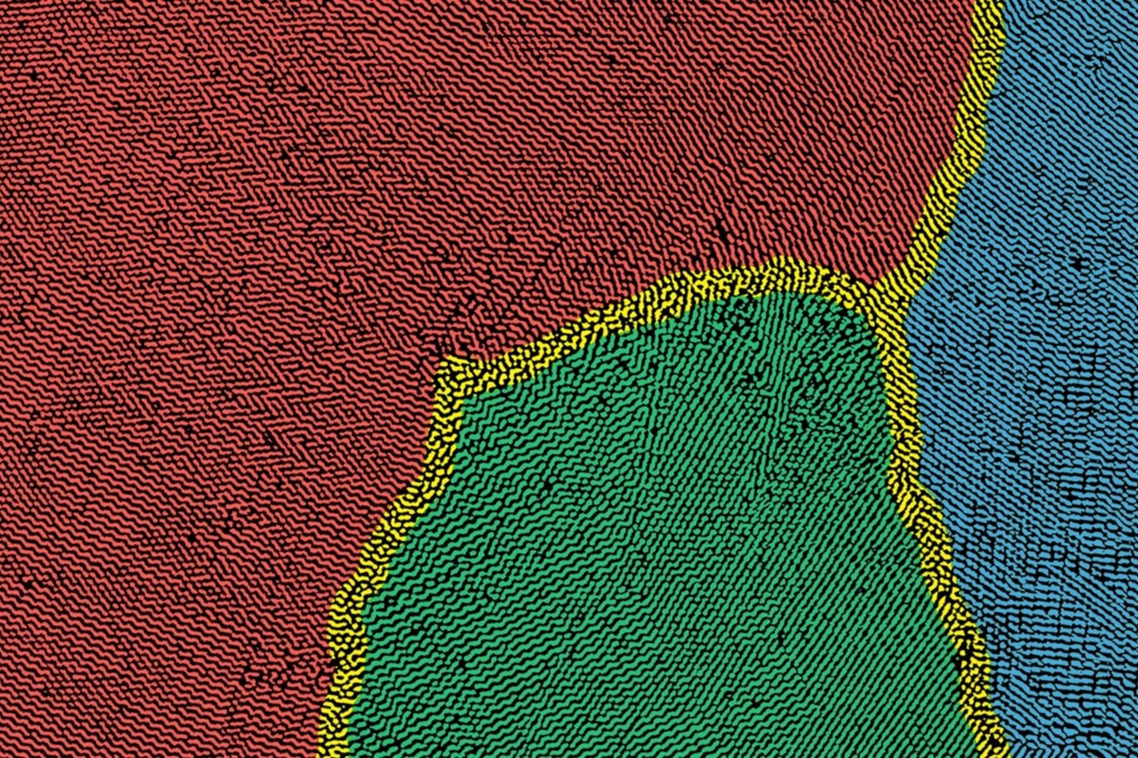
Nanoimaging Reveals Topological Textures in Nanoscale Crystalline Networks
X-ray nano-tomography reveals collective behavior in synthetic self-assembled nanostructures. The new method opens opportunities for the synthesis of photonic and plasmonic materials with improved long-range ordering.
Cause of clogged hypodermic needles discovered
Researchers at PSI and the ANAXAM technology transfer center have found the cause of clogging in prefilled syringes.
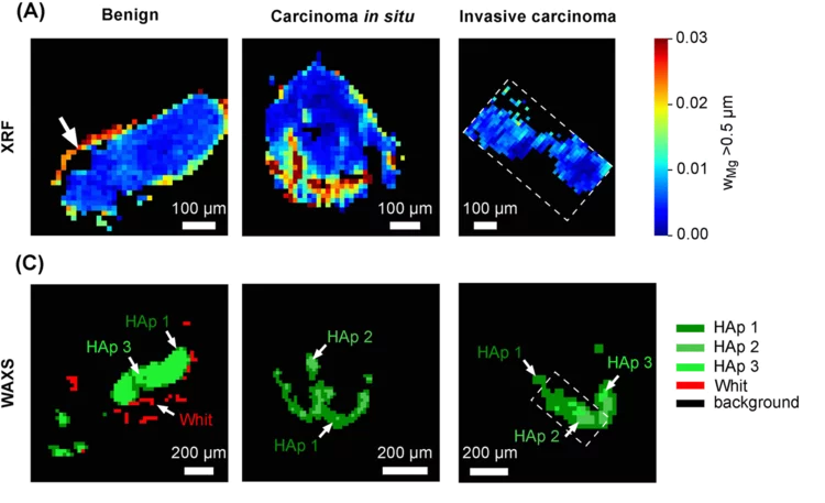
Whitlockite in mammary microcalcifications is not associated with breast cancer
Microcalcifications, small deposits of calcium-containing minerals that form in breast tissue, are often, but not always, a warning sign of breast cancer. The relationship between microcalcifications and cancer has not been fully understood thus far. Researchers discovered now that the relationship between microcalcifications and tumors seems to be linked to the presence of a particular mineral called whitlockite, which is rich in magnesium and is found in microcalcifications only in the absence of tumors.
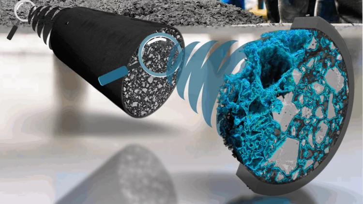
3D insights into an innovative manufacturing process
3D printing for creating complex shapes
Earlier detection of breast cancer
3D X-rays can improve breast cancer screening.
Bright white coloring of Pacific cleaner shrimp revelead
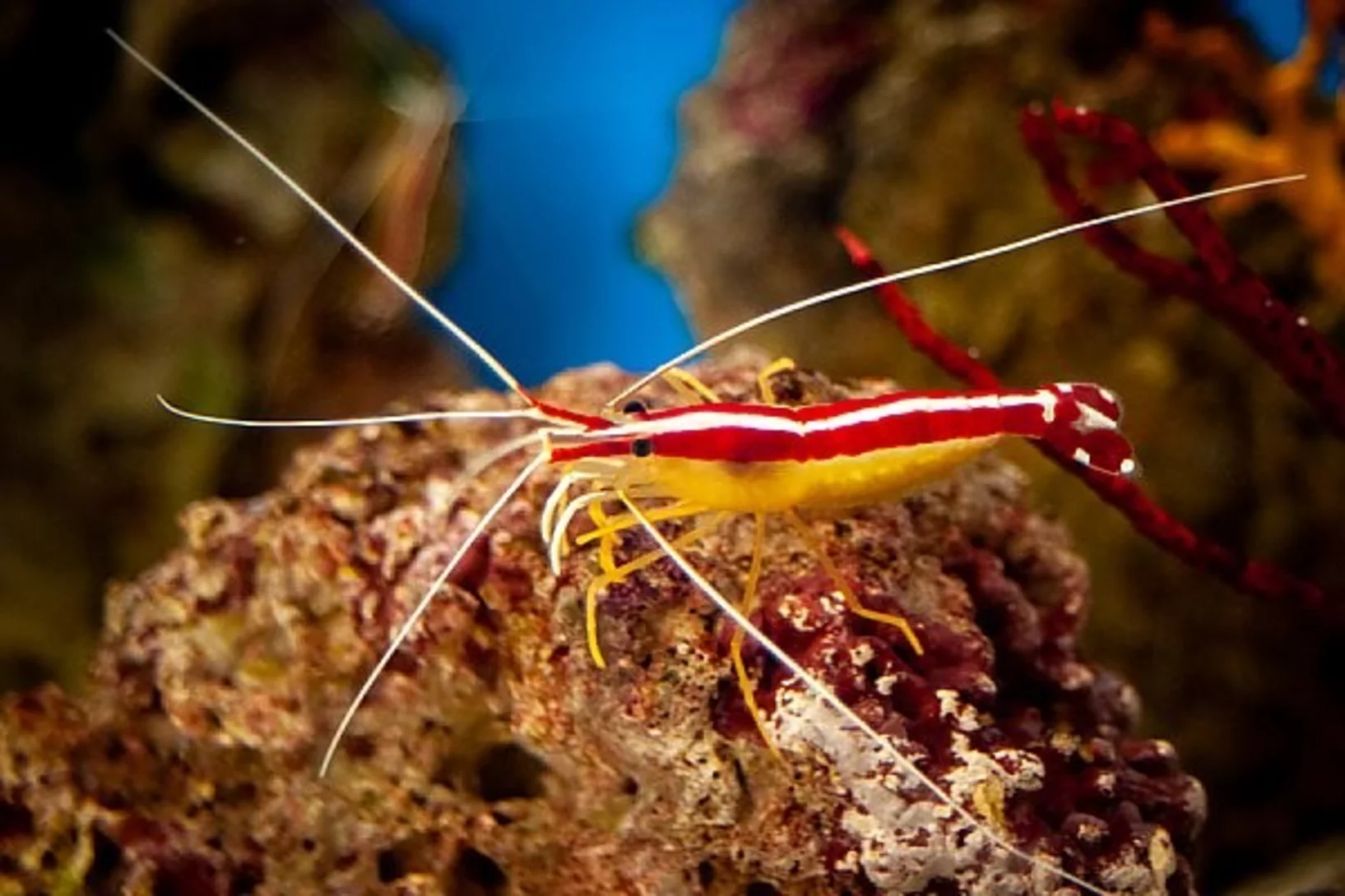
In a study published in Nature Photonics, researchers at the Ben-Gurion University of the Negev, Israel, explain the bright, white-colored stripes from Pacific cleaner shrimps, one of the most efficient white reflectors found in nature.
Progress of the X06DA-PXIII beamline upgrade: First light in the optics hutch
On June 7, 2023, the PXIII project team successfully shone the first light into the optics hutch at the upgraded X06DA-PXIII beamline. It is an essential first step for testing new hardware and software solutions that will be implemented at SLS2.0.
A deep look into hydration of cement
Researchers led by the University of Málaga show the Portland cement early age hydration with microscopic detail and high contrast between the components. This knowledge may contribute to more environmentally friendly manufacturing processes.
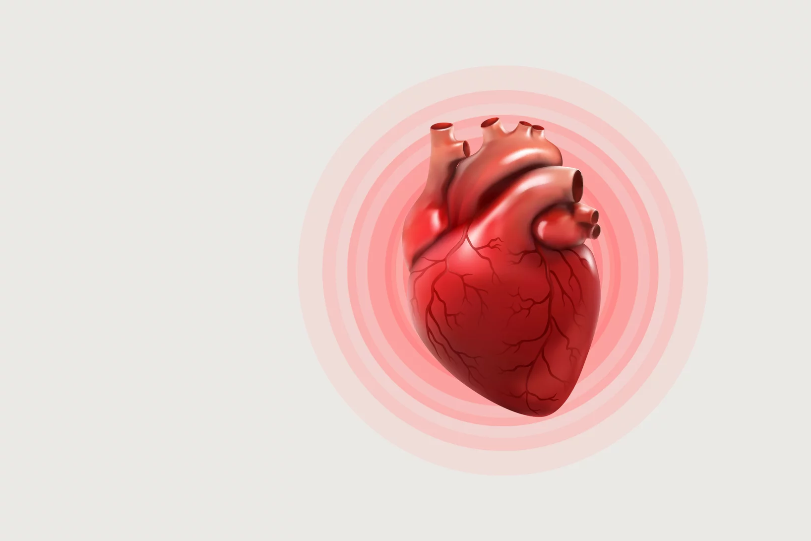
X-ray imaging after heart transplantations
Synchrotron light can be used in follow-up after a heart transplant to determine whether the body may be rejecting the new organ.
X-ray tomography helps understand how the heart beats
Researchers at the Swiss Light Source SLS use X-ray phase contrast imaging to study a heart in action as it beats.
Manuel Guizar-Sicairos appointed as Associate Professor at EPF Lausanne and head of the Computational X-ray Imaging group at PSI
Dr. Manuel Guizar-Sicairos, currently Senior Scientist at PSI, was appointed as Associate Professor of Physics in EPF Lausanne and head of the Computational X-ray Imaging group in PSI.
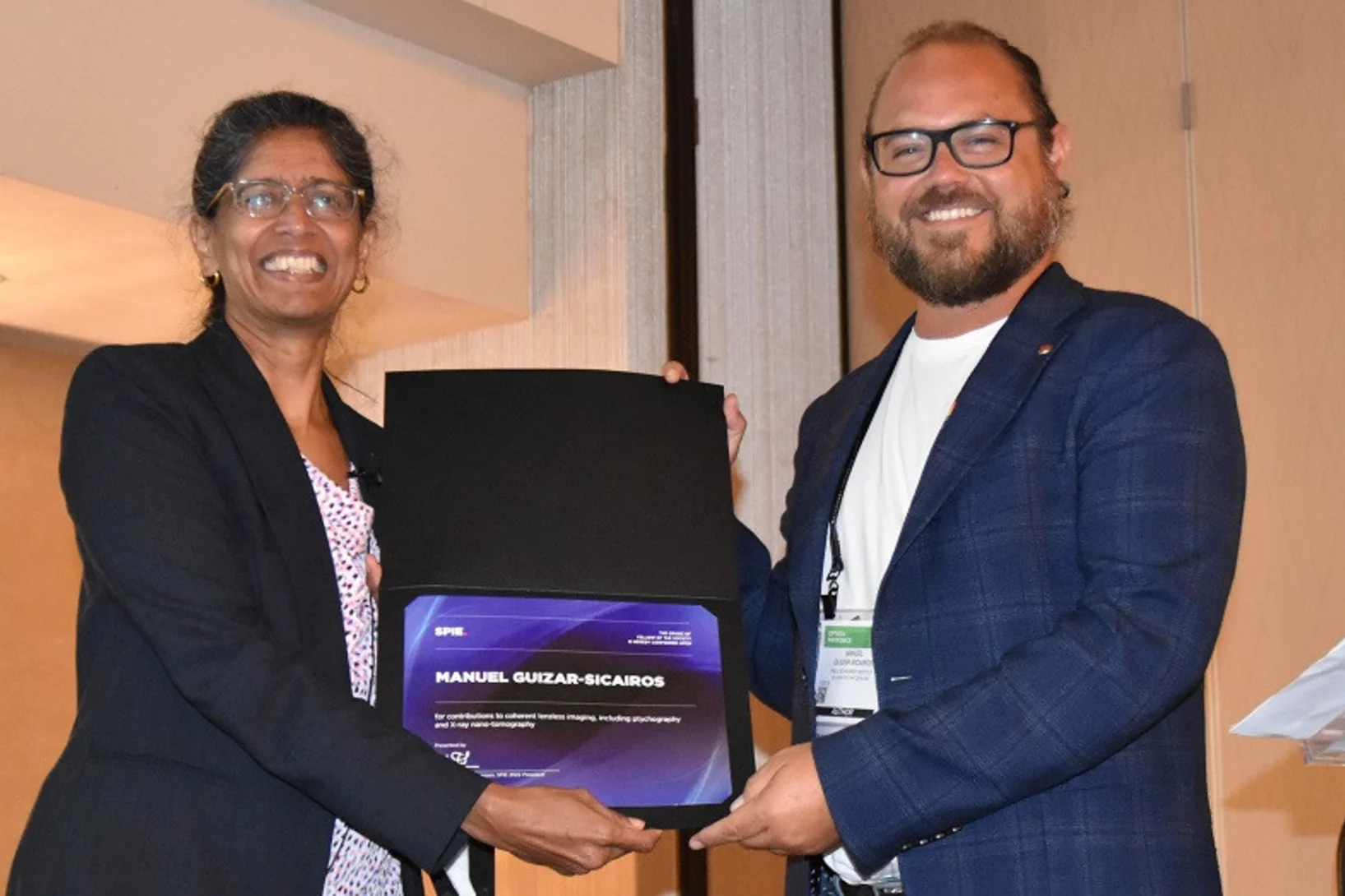
Dr. Manuel Guizar-Sicairos elected as SPIE Fellow member
Dr. Manuel Guizar-Sicairos was elected as a 2022 SPIE Fellow Member for his contributions to coherent lensless imaging, including ptychography and X-ray nano-tomography. The distinction was awarded in the SPIE’s Optics & Photonics conference in San Diego, California.
Nanomaterial from the Middle Ages
Unlocking the secrets of Zwischgold at PSI.
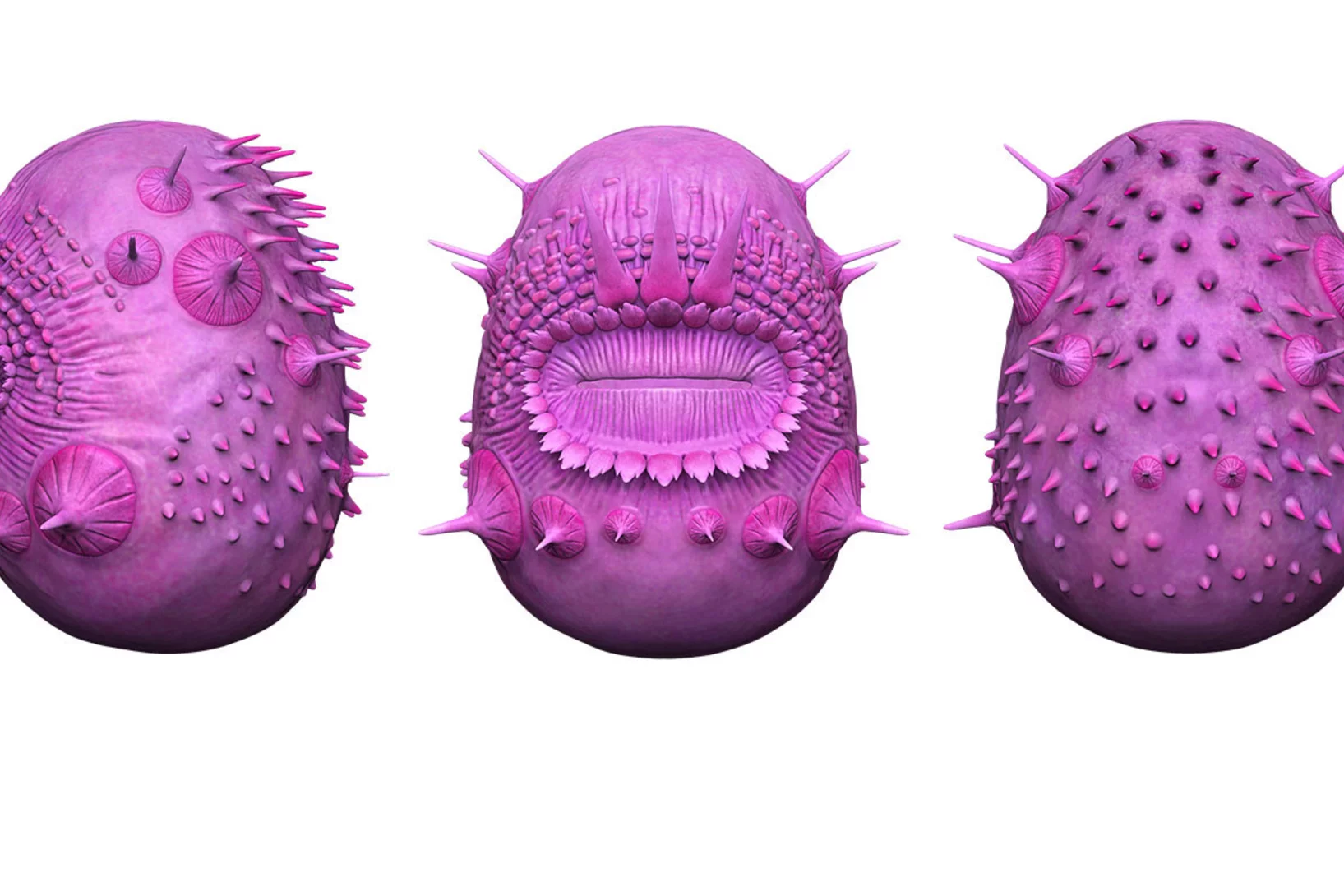
Weird fossil is not our ancestor
X-ray light solves puzzle of human ancestry
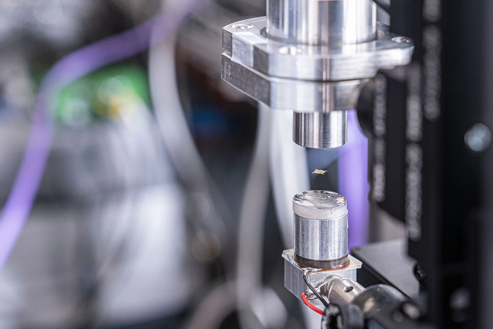
A new spin on sample delivery for membrane proteins
Proteins hover in front of the X-ray beam at a Swiss Light Source beamline. Now, spinning thin films bring on board these trickiest of proteins.
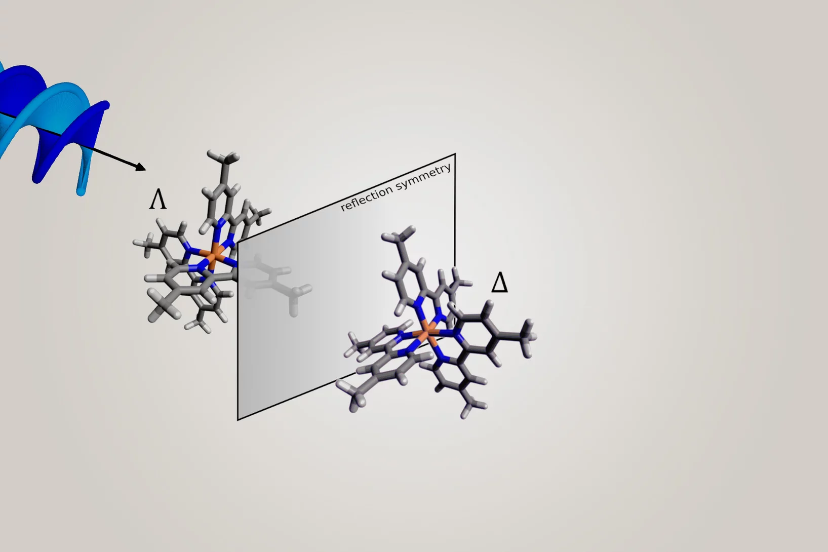
Making it easier to differentiate mirror-image molecules
Researchers have shown that mirror-image substances – so-called enantiomers – can be better distinguished using helical X-ray light.
Hercules School 2022
PSI hosted again the Hercules School in March 2022. We had the pleasure to welcome 20 international PhD students, PostDocs and scientists to demonstrate our state-of-the-art techniques and methodologies at our large scale facilities, the Swiss Light Source (SLS), the Swiss Spallation Neutron Source (SINQ) and our free electron laser SwissFEL.
Novel X-ray lens facilitates glimpse into the nanoworld
PSI develops a revolutionary achromatic lens for X-rays.
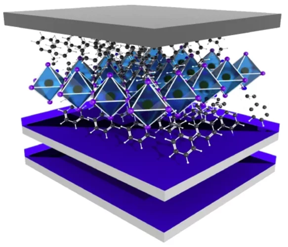
Lighting up the appealing world of hybrid perovskites
Researchers from Italy, in collaboration with the Paul Scherrer Institut, successfully used the macromolecular crystallography beamline X06DA-PXIII at the Swiss Light Source to characterize promising perovkites materials used in solar cells and other photodetector devices.
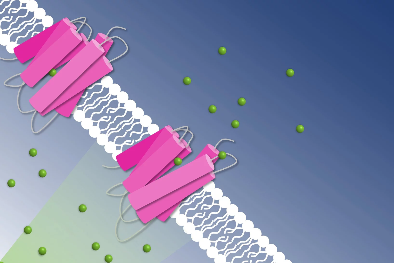
How to get chloride ions into the cell
A molecular movie shot at PSI reveals the mechanism of a light-driven chloride pump
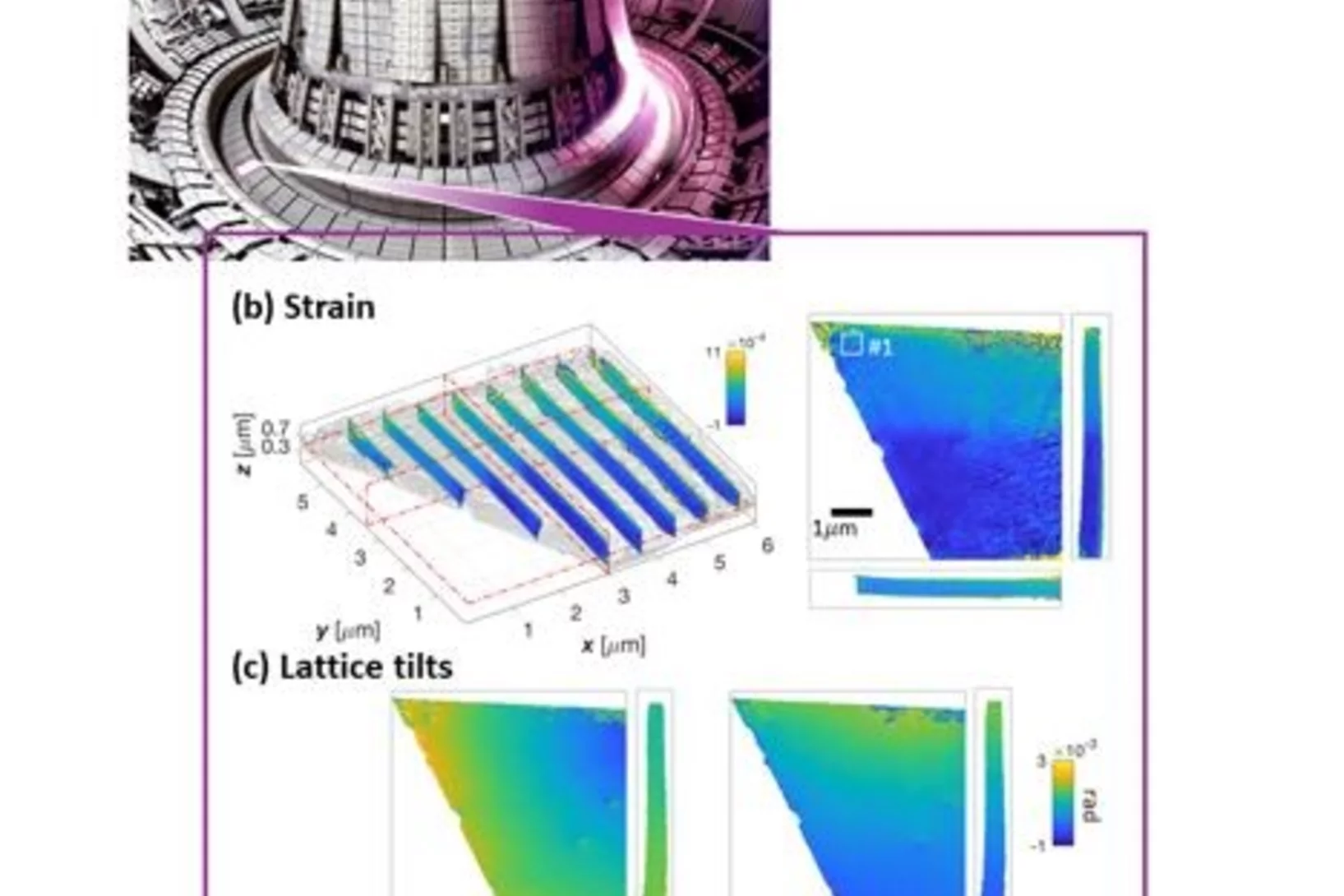
Revealing invisible defects in fusion reactor armor
In an exciting collaboration, Nick Phillips, a PSI Fellow at the cSAXS beamline, reveals nanoscale lattice distortions created by invisible defects in fusion reactor armor. This work develops the current understanding of how the smallest, but most prevalent defects, generated during neutron irradiation behave. The novel Bragg ptychographic approach published in Nature Communications paves the way for fast, robust, 3D Bragg ptychography.
Ground-breaking technology development recognised
PSI researchers win the international Innovation Award on Synchrotron Radiation for 3D mapping of nanoscopic details in macroscopic specimens, such as bone.
Glycation of collagen: Quantifying rates
Collagen is abundant in the connective tissue of human beings, e.g. in tendons, ligament and cornea. Glycation of collagen distorts its structure, renders the extracellular matrix stiff and brittle and at the same time lowers the degradation susceptibility thereby preventing renewal. Based on models and with parameters determined from experimental data, we describe the glycation of type 1 collagen in bovine pericardium derived bio-tissues upon incubation in glucose and ribose. We hope that this contributes to a better quantitative understanding of the effects of diabetes on collagen.
Dr. Manuel Guizar-Sicairos is awarded ICO prize
Dr. Manuel Guizar-Sicairos, beamline scientist at the cSAXS beamline, is the 2019 recipient of the International Commission for Optics (ICO) Prize. The distinction was awarded in the EOSAM conference in Rome.
Protein distancing
PSI researchers have developed a new method to attach proteins to the surface of virus-like particles.
X-ray microscopy with 1000 tomograms per second
A team at the Swiss Light Source SLS have set a new record using an imaging method called tomoscopy.
The mystery of the flexible shell
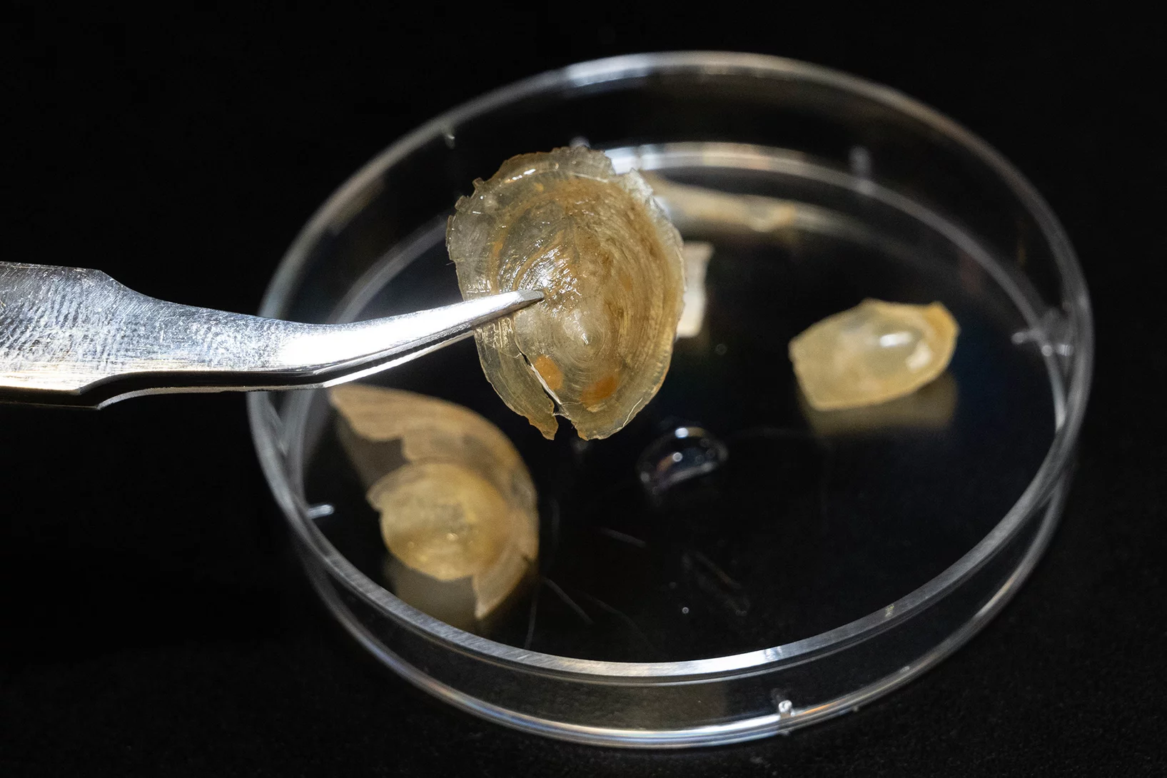
Why the shell of a marine animal is soft in water but hard in air.
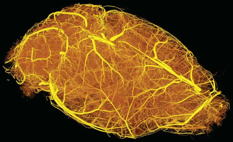
Hierarchical imaging and computational analysis of three-dimensional vascular network architecture in mouse brain
An international team involving researchers from the University and University Hospital Zürich, the Krembil Research Institute and the University and University Hospital in Toronto (Canada), the Department of Physics of Jyväskylä (Finland), the University of Leuven (Belgium), the Johannes Kepler University in Linz (Austria), the Novartis Institutes for Biomedical Research in Emeryville (USA), the ETH Zürich and the Paul Scherrer Institute has developed a protocol that enables hierarchical imaging and computational analysis of vascular networks in entire postnatal- and adult mouse brains, enabling direct and quantitative comparisons of the morphological brain vascular network architecture between different postnatal and / or adult developmental stages. The results have been published on Nature Protocols on September 3rd, 2021.


