Catalytic materials are ubiquitously used in industrial processes to perform chemical reactions efficiently and in a sustainable manner. Nanoporous gold (npAu) is a monolithic sponge-like catalyst exhibiting a hierarchical structure with pores and connecting ligaments of typically 10 to 50 nm. This greatly increases the fraction of active surface Au atoms available for chemical reactions compared to supported heterogeneous catalysts, and makes npAu interesting for a variety of catalytic and sensing processes. Due to the highly flexible structure of npAu, including variable pore/ligament size and presence of dopants or bimetallic species, it is essential to develop a deep understanding of catalytic performance by deriving structure-activity relationships. This in turn leads to the rational design of more efficient catalysts tuned for specific reaction conditions. The ultimate goal is to visualize catalyst structures in three dimensions (3D) in situ, i.e. under real reaction conditions.
Electron microscopy is the most established approach for high resolution visualization of materials. However, while even sub-nm analysis is routinely available, the low penetration depth of electrons rapidly becomes a limiting factor. In the case of npAu catalytic specimens, electron tomography (ET) can be performed on slices of material about 300 nm thick with a few nm resolution, which allows 3D structural visualization with great detail. However, the high vacuum environment makes ET very challenging to perform under real conditions. Moreover, the reduced sample size may lead to non-representative sampling of the diverse hierarchical structure.
Alternatively, serial sectioning with a focused ion beam (FIB) combined with surface analysis by scanning electron microscopy (SEM) allows probing large volumes of material of up to a few tens of microns with a resolution below 15 nm. However, high vacuum conditions are also required, while the sample is completely destroyed during imaging, which prevents repeated imaging on the very same specimen.
In the context of catalysis where material structure is intrinsically related to chemical function, X-ray tomography is an ideal technique for imaging catalysts in 3D under real working conditions, while preserving structural integrity. Due to the penetration power of hard X-rays, large sample volumes can be probed at atmospheric or elevated pressure and temperature conditions non-destructively, which allows comparative imaging after different catalytic processes. What remains a challenge in X-ray imaging is approaching resolutions on the nanometer length scale.
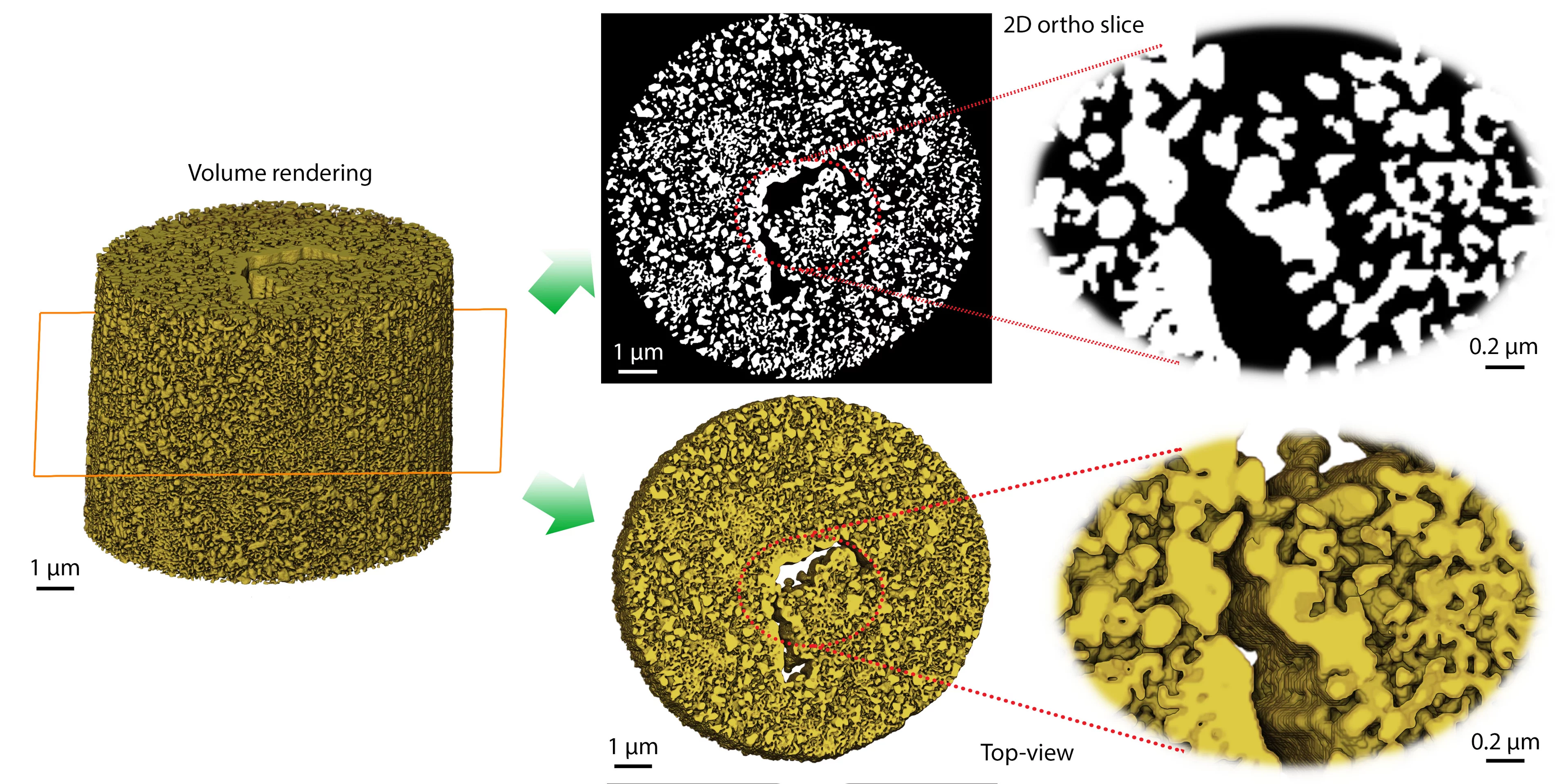
Researchers from Karlsruhe Institute of Technology and the University of Bremen are exploring techniques that will enable them to pursue their goal: characterizing the extended hierarchically-porous structure of a npAu of a few microns in size during or after catalytic operation. For this purpose they have performed ptychographic X-ray computed tomography (PXCT) at the cSAXS beamline at the Swiss Light Source, using the highly-innovative flOMNI instrument, which makes use of laser interferometric metrology for high-accuracy scanning of the sample. In a proof-of-principle experiment, a fragment of about 10×10×10 µm3 was imaged in 3D with very high resolution. Although it is difficult to estimate the spatial resolution with high accuracy, their analysis has shown that features in the sample as small as 23 nm are correctly reproduced.
Although electron microscopy techniques currently provide higher resolution, the image quality by PXCT reported in this work will enable to follow coarsening effects in a representative volume of the sample sequentially after thermal annealing. This is planned for future experiments with which researchers aim to investigate the stabilization of npAu catalysts at high temperatures, potentially leading to improvements in performance and stability during selective oxidation reactions.
Contact
Dr. Ana diazBeamline Scientist, Swiss Light Source
Paul Scherrer Institut
Telephone: +41 56 310 5626
E-mail: ana.diaz@psi.ch
Original Publication
Correlative Multiscale 3D Imaging of a Hierarchical Nanoporous Gold Catalyst by Electron, Ion and X-ray NanotomographyY. Fam, T. L. Sheppard, A. Diaz, T. Scherer, M. Holler, W. Wang, D. Wang, P. Brenner, A. Wittstock, and J.-D. Grunwaldt
ChemCatChem 10 2858 (2018)
DOI: 10.1002/cctc.201800230
