
In a new study on retinal degeneration linked to blindness, a team of collaborators from the photon-science and biology divisions of PSI, University of Zürich, and ETHZ, could see structures inside the nerve cells of the retina of a mouse. Biological tissue samples were imaged using ptychographic X-ray computer tomography (PXCT) at a resolution which allows to follow in three dimensions the axons path (blue arrow) of the retina in 3D. The technique allows to observe subcellular elements of the synapses (orange arrow) like the large mitochondria and the specialized ribbon responsible for neurotransmission, at an isotropic resolution of 150-200 nm.
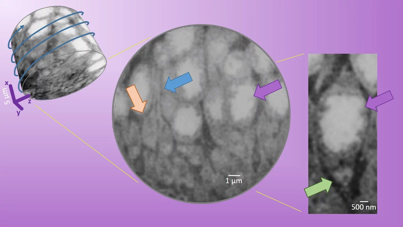
Retinas from models of neurodegenerative disease were compared to the wild-type tissue. In the transgenic mouse model of retinitis pigmentosa, most synapses and axonal fibers of the photoreceptor cells are absent, and fibrosis prevails. The tomograms reveal the arrangement of more than 60 cell bodies in a cylindrical volume of 25 µm diameter and 20 µm height (upper left in figure 1). Because PXCT is a non-destructive technique, the researchers could perform complementary analysis of the sample from a healthy mouse at higher resolution with sequential nanometer slicing using a focused-ion beam and imaging with a scanning electron microscope. X-ray nanotomography showed a good match of the subcellular structures like the ribbon synapse and even a somatic synapse.
The study paves the way for correlative analysis of neural tissues subject to neurodegeneration, remodeling and blindness treatments.
The work is online here and is selected as Research Highlight: Non-destructive imaging of large tissue volumes using X-rays
Read the full story
Imaging of retina cellular and subcellular structures using ptychographic hard X-ray tomography.
Panneels V, Diaz A, Imsand C, Guizar-Sicairos M, Müller E, Bittermann AG, Ishikawa T, Menzel A, Kaech A, Holler M, Grimm C, Schertler G.
J Cell Sci. 2021 134(19):jcs258561. doi: 10.1242/jcs.258561. Epub 2021 Oct 14. PMID: 34494099
Contact
Dr. Ana Diaz,
Swiss Light Source
Paul Scherrer Institut, 5232 Villigen PSI, Switzerland
Phone: +41 56 310 5626, e-mail: ana.diaz@psi.ch
https://www.psi.ch/en/sls/csaxs
Dr. Valérie Panneels,
Laboratory of Biomolecular Research
Paul Scherrer Institut, 5232 Villigen PSI, Switzerland
Phone: +41 56 310 2104, e-mail: valerie.panneels@psi.ch
https://www.psi.ch/en/lbr