

The PSI Center for Life Sciences is very well equipped for imaging both live and fixed specimen of cells and tissues. Our microscopes are used to monitor subcellular localization of proteins and for the functional analysis of cellular processes using for instance GFP tagged proteins.
The light microscopy facility operated by the MGG group at LNB includes the following instruments:
This microscope can be used for widefield and confocal imaging. Laserlines at 405, 488, 561, 640 nm and filtersets for DAPI, CFP, FITC, YFP, TRITC, mCherry, Cy5, and Cy7. It has objectives from 10x to 100x.
STELLARIS 5 LIAchroic is a true confocal point scanning system with LIAchroic beam splitters. It includes a highly sensitive, prism-based spectral detection design with computer controlled adjustable bandwidth for all fluorescence channels. Laserlines at 405, 488, 561, and 638 nm, Detection range 410-850 nm. It is equipped with 20x and 63x objectives.
The IXplore TIRF microscope system offers simultaneous multicolor TIRF imaging for up to 4 colors. Laserlines at 405, 488, 561, and 640 nm. Objectives from 20x to 100x including a special TIRF objective. It is possible to operate it between 15 and 37 degree for life cell imaging.
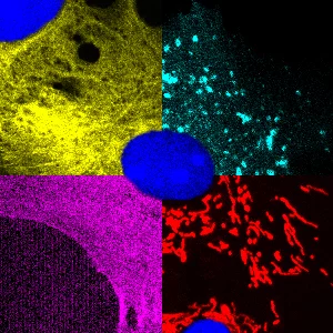
Leica SP5 Confocal Microscope
This confocal microscope is equipped with three lasers providing laser lines from 458nm to 633nm. This allows us to separate up to four fluorescent proteins or dyes. It is possible to operate it between 15 and 37 degree. It is located at the Center for Radiopharmaceutical Sciences.