We develop novel methods for visualizing life at atomic resolution using electron diffraction and coherent electron diffractive imaging
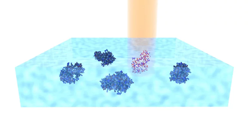
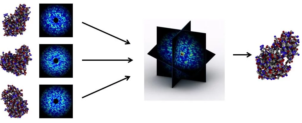
To comprehend how matter creates life requires visualising the living cell in atomic detail. To this aim, we develop novel technologies that are based on electron diffraction of frozen, hydrated biological samples. We have demonstrated (using biological and organic nano-crystals) that electron diffraction data can in principle unlock high-resolution information way beyond what conventional electron microscopy can deliver, and that this also applies to other biological samples. To exploit this novel approach in the study of cellular processes, we scan samples with a coherent beam with a diameter down to several nanometers, and measure the diffracted electrons using modern hybrid pixel detectors as developed by PSI’s detector group. For phasing of these data, we develop a range of methods that combine experimental and computational approaches. We apply our research to the study of the molecular response to mitochondrial stress central to neuro-degeneration and aging.


