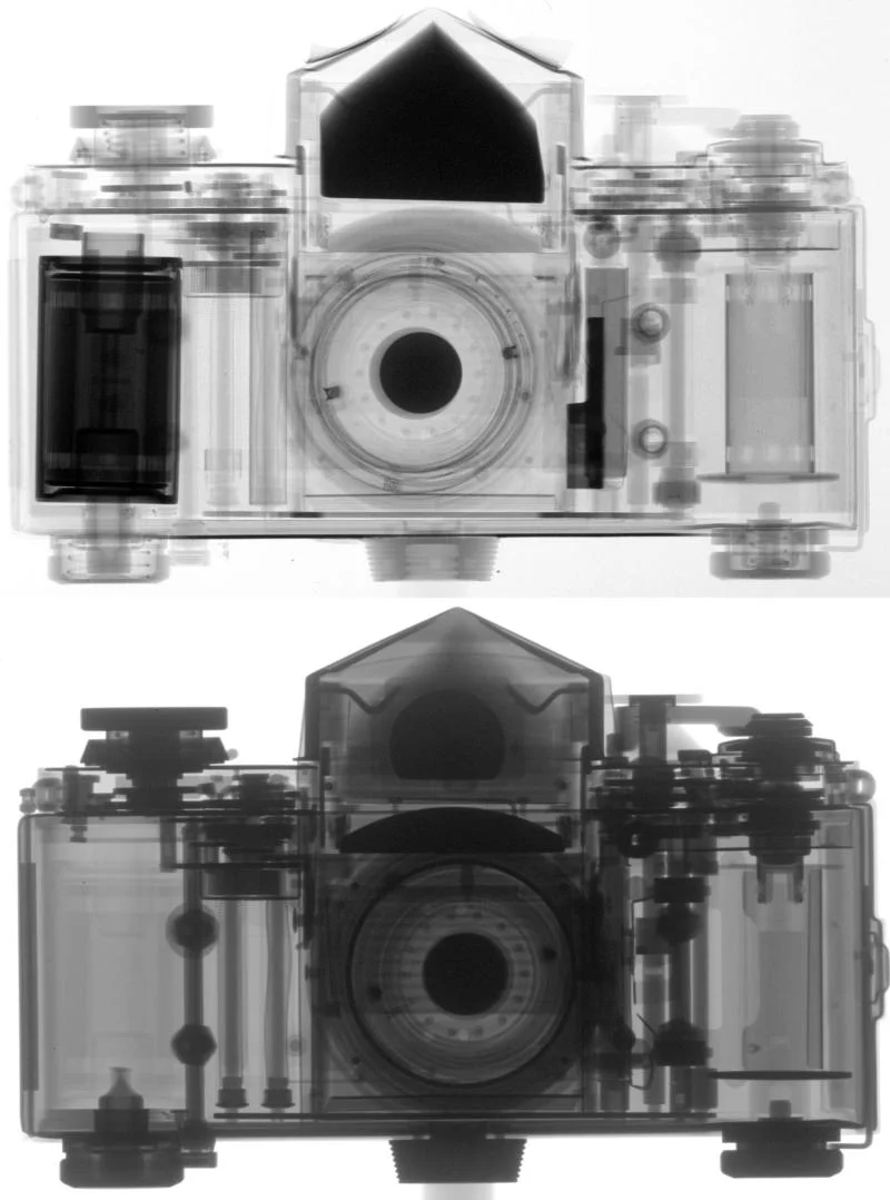
Neutron Imaging (NI) is a radiographic testing method using neutrons. As such it is quite similar to X-ray, which is well known from medical applications. Neutrons are able to transmit material layers of certain thickness which depends on the specific attenuation properties of that material. This enables to establish neutron imaging as a non-destructive inspection method which can favorably be applied for research and practical, industrial related problems. A similarity and complementarities to the conventional X-ray imaging are given. Because different materials have different attenuation behaviour the neutron beam passing through a sample can be interpreted as signal carrying information about the composition and structure of the sample.
Learn more about neutron imaging from the PSI Neutron Imaging Brochure or from the sections:


