
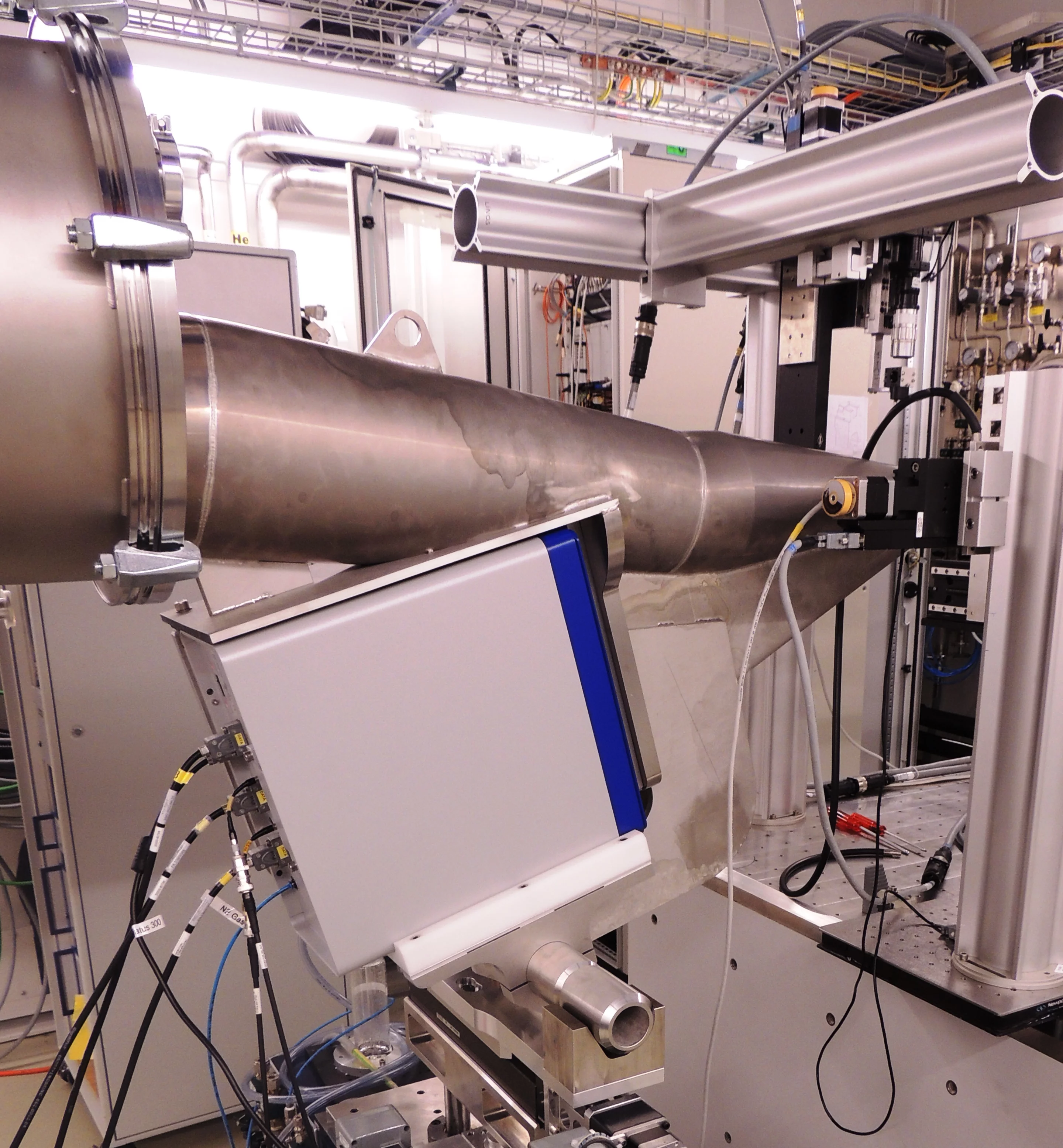
Two evacuated flight tubes are available, 7m and 2.1m long, respectively, in order to offer flexibility for a wide range of reciprocal- and/or real-space requirements. The flight tubes have a small entrance mica window of about 7 micron thickness, and a 300 micron-thick exit mylar window matching the size of the Pilatus 2M detector. For some experiments it is beneficial to have a thinner exit window and for these cases we can flush these flight tubes with Helium and use a thin mylar exit window. Note that no interchange of flight tubes will be practical during your beamtime. Please discuss with the beamline staff ahead of time, which geometry will be practical for your experiments.
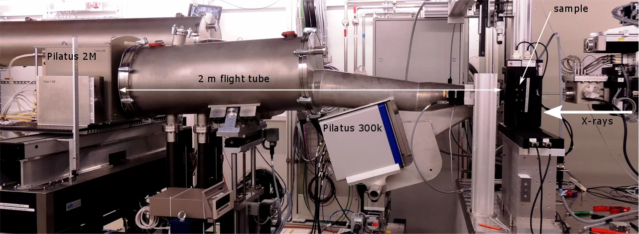
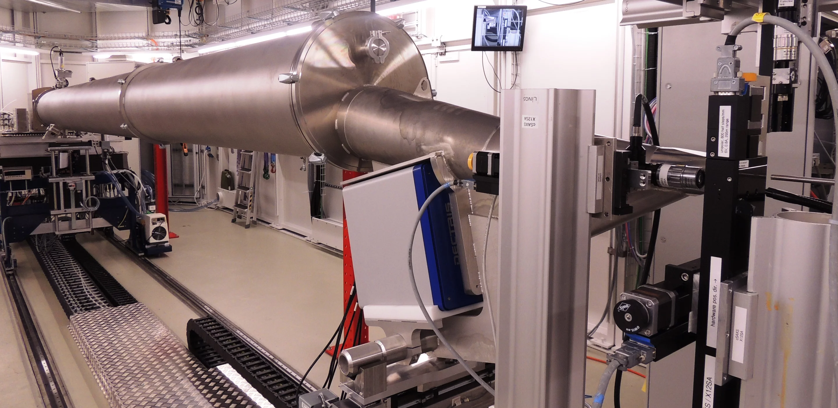
The 7m and 2.1m long flight tubes allow for combined SAXS/WAXS measurements. The WAXS signal is currently measured with a Pilatus 300k. Notice the detector is oriented as a vertically oriented strip, no orientation information from anisotropic scattering is then available for the WAXS detector in this configuration.
cSAXS is a beamline dedicated to a variety of techniques, and experiments are performed on a wide assortment of samples. Consequently, there is no "typical" experimental setup. Instead, for each user group, a set up is custom built. Therefore the experiment should be discussed as early as possible with the beamline staff. We encourage users to come at least a day in advance so that building and organizing the sample environment, as well as special detector needs, can be realized before the beamtime starts.
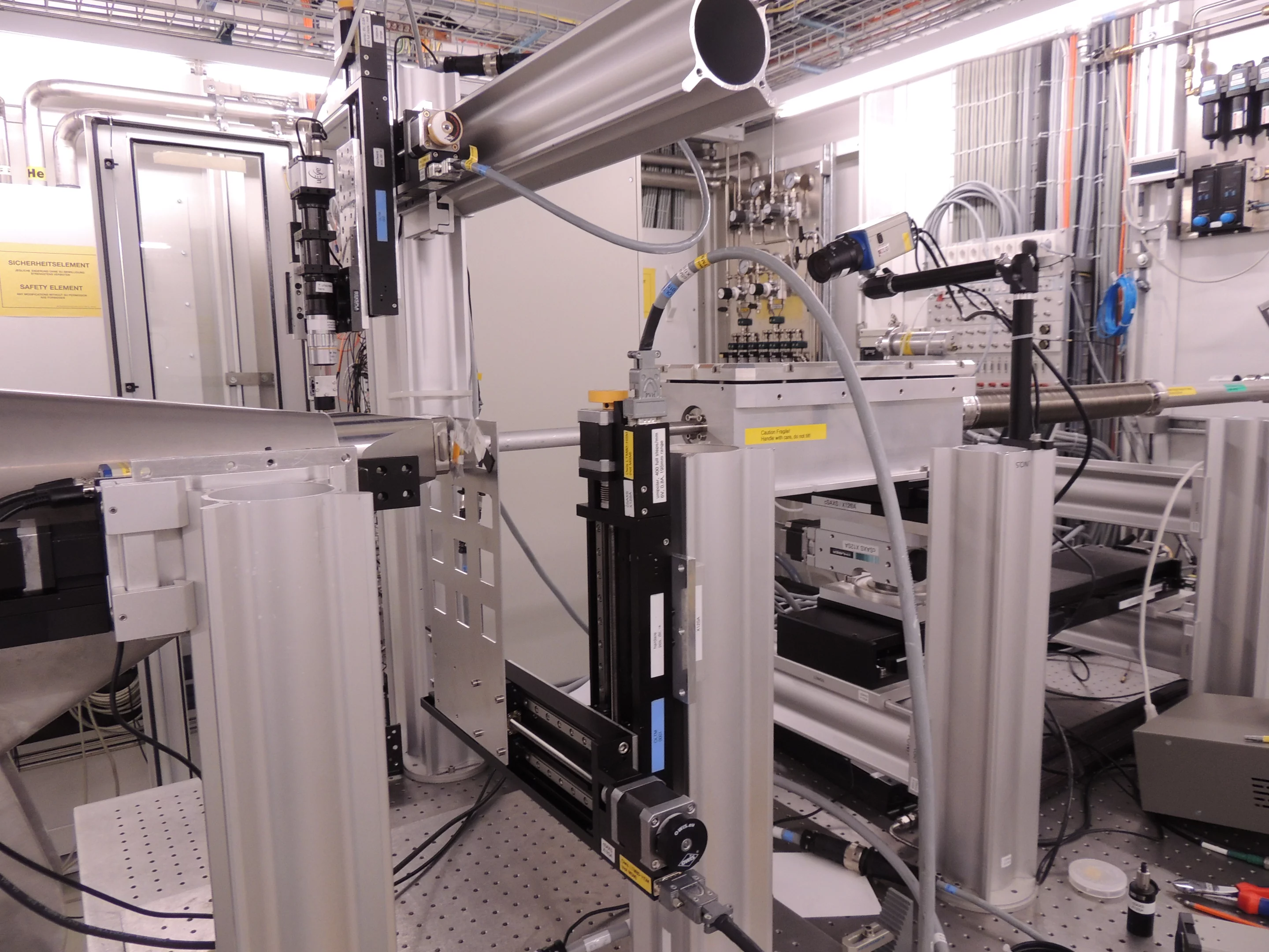
Common to essentially all experiments is that the beam passes approximately 550 mm above an optical table, which serves as sample table. We have a variety of Al-profiles, rails, motorized stages, etc., which can be assembled as needed. All of these stages are interfaced with spec, the control software at the beamline.
Currently, we do not support a completely evacuated beam path/sample environment.
In the following we list semi-standard setups.
Scanning SAXS in 2D
Scanning SAXS bridges the gap between high resolution local probes and low resolution techniques that are capable of imaging larger sample areas. The sample is raster scanned through the beam. While moving at constant speed along a line of this raster scan the detector records a series of 2D SAXS data. In this way typically a few ten thousand SAXS images are recorded per hour and the scattering technique SAXS can be used for various imaging modes. Nanoscale features, averaged over an X-ray spot size on the order of 100 μm2, are thereby imaged over square millimeter or centimeter sized areas. See for example Bunk et al., New J. Phys. 11, 123016 (2009).
Scanning SAXS in 3D
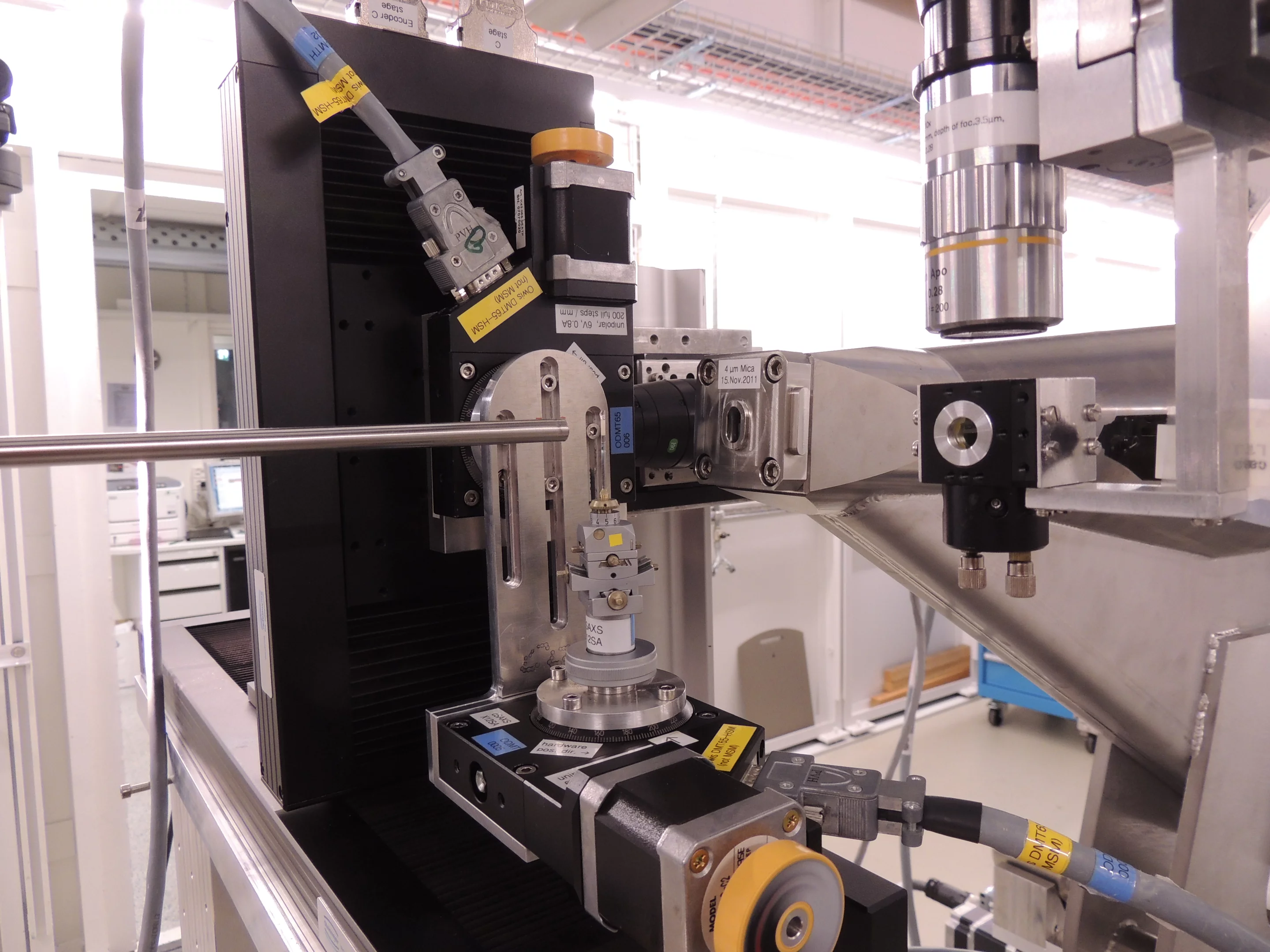
Combining scanning SAXS with two axes of rotation enables SAXS tensor tomography. With this technique one can obtain the 3D orientation of nanostructures in a bulk specimen spatially resolved in 3D. An example can be found in Liebi et al., Nature 527, 349 (2015). Reciprocal space information is given by the q-range available to the SAXS detectors and real-space resolution is given by the size of the beam, at cSAXS it is possible to use this technique with a resolution between 2 and 500 microns.
Ptychography in 2D
Ptychography is a high resolution scanning microscopy technique based in coherent diffractive imaging where the resolution is not limited by the spot size of the X-ray beam. At cSAXS resolutions down to 8 nm can be reached for strongly scattering samples. but one should keep in mind that resolution is limited by the sample scattering. Typical scan areas are on the order of a few 10 μm2. See Vila-Comamala et al., Opt. Express 19, 21333 (2010) for an example.
Ptychographic X-ray Computed Tomography (PXCT)
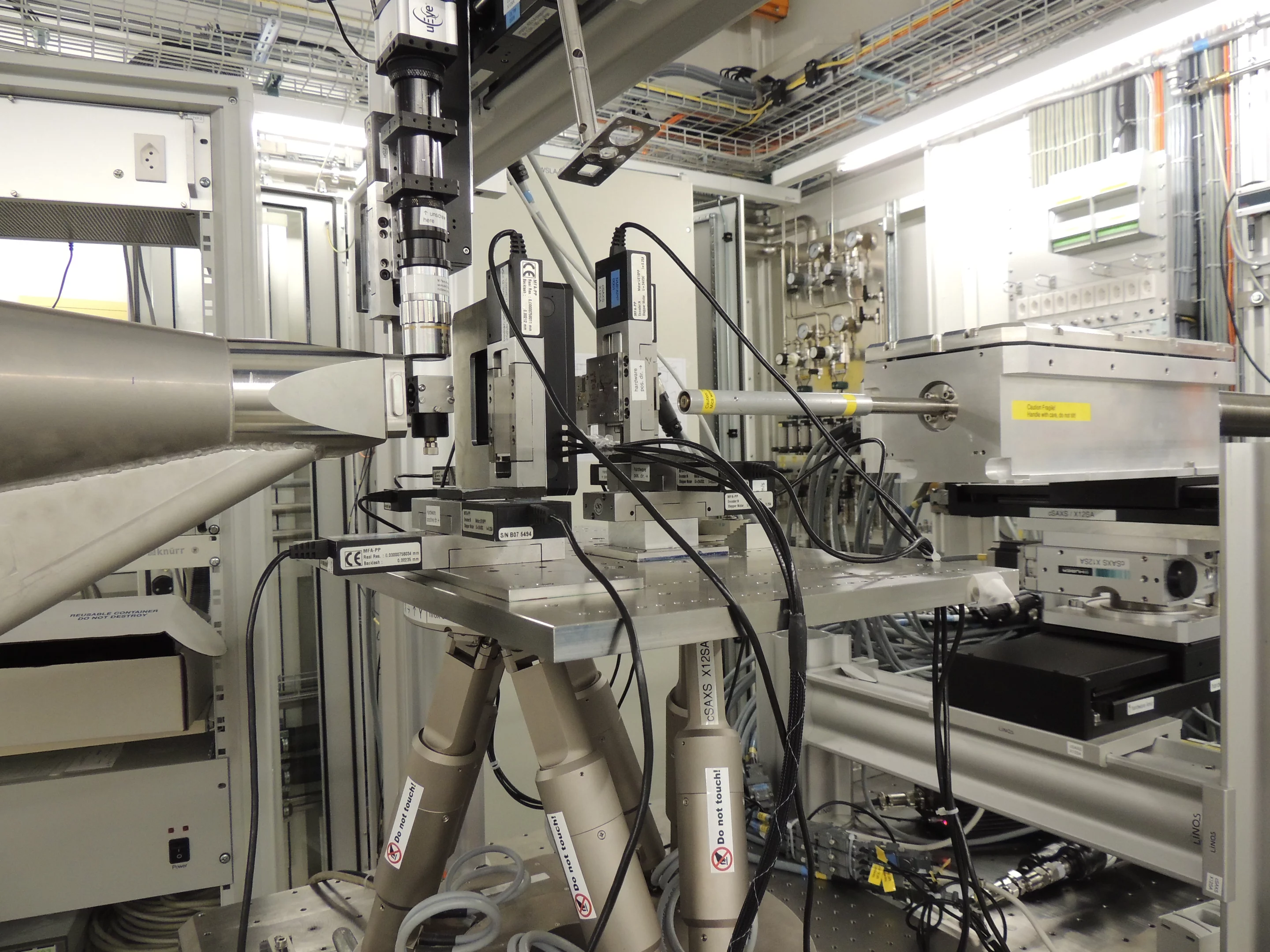
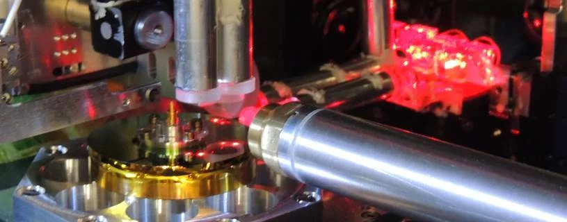
Adding a rotation degree of freedom to ptychography enables high-resolution tomography, see for example Dierolf et al., Nature 467, 436-439 (2010). For a spatial resolution below 100 nm in 3D, each 2D projection must be recorded without any distortion, for which accurate positioning of the specimen with respect to the illumination beam is required. The instruments described below, flOMNI and OMNY, use differential laser interferometry for an accurate positioning of the sample with respect to the beam.
Specimens for these instruments have particular requirements, you can read some general guidelines below.
flOMNI - A flexible instrument for PXCT
This instrument operates at atmospheric pressure and at room temperature, which limits its use to materials that are not radiation sensitive. In turn this instrument allows for more flexibility and customization on the sample environment. For example to provide to the sample electrical or mechanical stimuli, or variations in temperature or gas. PXCT with a resolution of 16 nm in 3D has been demonstrated in M. Holler et al., Sci. Rep. 4 3857, (2014) and a resolution of 20 nm is feasible with strongly scattering samples.
OMNY - An instrument for cryo PXCT
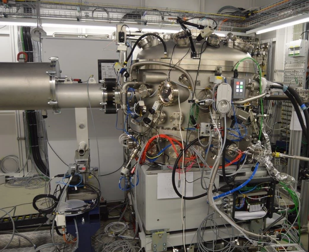
This instrument makes use of an identical laser interferometer concept for accurate positioning of the sample with respect to the illumination, but it additionally has a cryo-stage at 90 K and is operated in ultra-high vacuum. Therefore, this instrument is ideal for samples that require cryogenic protection to radiation damage, or for cryogenically fixed specimens. The OMNY is equipped with a sample transfer unit such that cryogenically prepared specimens can be transferred in cryogenic conditions.
Sample preparation for flOMNI and OMNY
Specimens for flOMNI and OMNY must be mounted on a special sample mount, please see Holler et. al. Rev. Sci. Instr. 88, 113701 (2017) and contact the beamline staff for questions or more details.
In order to perform high-resolution ptychographic tomography the entire sample volume needs to be confined within a cylinder with a maximum diameter of about 100 microns, such that there is air at both sides of sample for all rotation angles from 0 to 180 degrees. This can be achieved by using focused ion beam (FIB) to cut a portion of a bulk sample, or by confining it in a glass microcapillary.
The resolution in ptychography is limited by the size and density contrast of the features in the sample and by the dose. In practice the scanning time is a key consideration based on the measured volume and sought resolution. As an example, a 20 micron diameter sample with 20 micron height with small and strong contrast features can be currently scanned in about 24 hours aiming for 20 nm resolution or in 1.5 hours for 40 nm resolution. We also offer a version of nearfield ptychography that allows for example scanning a 100 micron diameter sample, with 35 micron height, in 5 hours aiming for 100 nm resolution. Scanning time scales linearly with volume and inversely to the fourth power of the resolution.
Time Resolved SAXS
Time resolved measurements in the millisecond range can be performed with the PILATUS and EIGER pixel detectors. Possible experiments include structural changes induced by laser radiation, microfluidic mixing or temperature changes. These SAXS or WAXS experiments will be often quite specific and it is important to contact beamline staff as early as possible, ideally before submitting a proposal for beamtime.
The following detectors are available at cSAXS:
| Type | Description | Array Size | Pixel Size | Active Area |
|---|---|---|---|---|
| PILATUS 2M | A PSI-developed detector, which operates in single-photon counting mode and thus allows operation without read-out noise, 20bit dynamic range. Continuous acquisition rate up to 10Hz, in burst mode 30Hz for ~2min For faster readout, up to ~80Hz, and reduced data volume you can choose to readout only the central 2×3 modules or, even faster, up to ~150Hz, the central 2 modules. |
1475×1679 pixels | 172×172 µm2 | 254×289 mm2 |
| 300k-W PILATUS | A three module PILATUS for up to 300Hz acquisition rate | 1475×195 pixels | 172×172 µm2 | 254×34 mm2 |
| 100k PILATUS (only upon request well in advance) | A single 100k PILATUS for up to 300Hz acquisition rate | 487×195 pixels | 172×172 µm2 | 84×34 mm2 |
| 500k Eiger | PSI-developed detector, single-photon counting, 4 to 32 bit dynamic range. Continuous acquisition rate up to 20 Hz or 60Hz depending on bit dynamic range, for rates exceeding the data transfer currently available the maximum number of frames that can be stored on the detector depends on the bit mode. Please contact the beamline staff for details. | 512×1024 pixels | 75×75 µm2 | 84×34 mm2 |
| Oxford Danfysik CyberStar | - | 15mm | - | |
| Amptek XR100-CR | An energy-dispersive detector. Si thickness = 500µm. |
- | 6 mm2 | - |
An Eiger 9M and an Eiger 1.5M will replace our current PILATUS 2M and PILATUS 300k for SAXS and WAXS measurements in the coming months. These detectors will have a very high frame-rate capability. For technical details and an update of when the detectors will be available please contact the beamline staff.
Several publications describe PILATUS and Eiger technology, see e.g.,
- B. Henrich B, A. Bergamaschi, C. Broennimann, R. Dinapoli, E.F. Eikenberry, I. Johnson, M. Kobas, P. Kraft, A. Mozzanica, and B. Schmitt, PILATUS: A single photon counting pixel detector for X-ray applications, Nucl. Instrum. Methods Phys. Res. A 607 247-249 (2009).
DOI: 10.1016/j.nima.2009.03.200 - R. Dinapoli, A. Bergamaschi, B. Henrich, R. Horisberger, I. Johnson, A. Mozzanica, E. Schmid, B. Schmitt, A. Schreiber, X. Shi, and G. Theidel, Eiger: Next generation single photon counting detector for x-ray applications, Nucl. Instr. Meth. Phys. Res. A 650, 79–83 (2011).
DOI: 10.1016/j.nima.2010.12.005. - I. Johnson, A. Bergamaschi, H. Billich, S. Cartier, R. Dinapoli, D. Greiffenberg, M. Guizar-Sicairos, B. Henrich, J. Jungmann, D. Mezza, A. Mozzanica, B. Schmitt, X. Shi, and G. Tinti, Eiger: a single-photon counting x-ray detector, J. Instrum. 9, C05032 (2014).
DOI: 10.1088/1748-0221/9/05/C05032.