Ferrous Iron Formation Following the Co-aggregation of Ferric Iron and the Alzheimer's Disease Peptide β-amyloid (1–42)
Everett J, Céspedes E, Shelford LR, Exley C, Collingwood JF, Dobson J, van der Laan G, Jenkins CA, Arenholz E and Telling ND
Journal OF THE ROYAL SOCIETY: INTERFACE 11, 20140165 (2014).
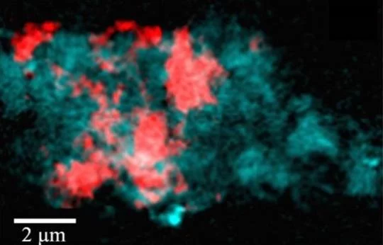
For decades, a link between increased levels of iron and areas of Alzheimer's disease (AD) pathology has been recognized, including AD lesions comprised of the peptide β-amyloid (Aβ). Despite many observations of this association, the relationship between Aβ and iron is poorly understood. Using X-ray microspectroscopy, X-ray absorption spectroscopy, electron microscopy and spectrophotometric iron(II) quantification techniques, we examine the interaction between Aβ(1–42) and synthetic iron(III), reminiscent of ferric iron stores in the brain. We report Aβ to be capable of accumulating iron(III) within amyloid aggregates, with this process resulting in Aβ-mediated reduction of iron(III) to a redox-active iron(II) phase. Additionally, we show that the presence of aluminium increases the reductive capacity of Aβ, enabling the redox cycling of the iron. These results demonstrate the ability of Aβ to accumulate iron, offering an explanation for previously observed local increases in iron concentration associated with AD lesions. Furthermore, the ability of iron to form redox-active iron phases from ferric precursors provides an origin both for the redox-active iron previously witnessed in AD tissue, and the increased levels of oxidative stress characteristic of AD. These interactions between Aβ and iron deliver valuable insights into the process of AD progression, which may ultimately provide targets for disease therapies.
Press Release: Microscopy & Analysis
Sub-25 nm direct write (maskless) X-ray nanolithography
Leontowich AFG, Hitchcock AP, Watts B and Raabe J
MICROELECTRONIC ENGINEERING 108, 5–7 (2013).
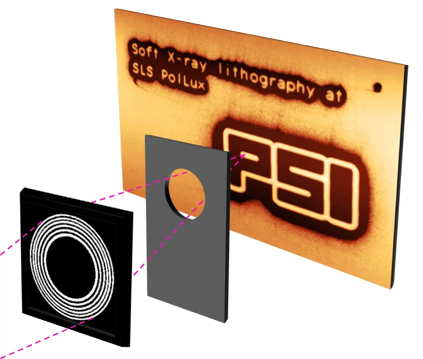
Sub-25 nm continuous, reproducible features of arbitrary geometry were created by X-ray lithography in direct write (maskless) geometry, analogous to conventional electron beam lithography. This was achieved through the use of a laser interferometer-controlled scanning transmission X-ray microscope (STXM) equipped with a zone doubled Fresnel zone plate lens, and a cold development procedure. These features are among the smallest created with photons in direct write geometry.
Direct Observation of Antiferromagnetically Oriented Spin Vortex States in Magnetic Multilayer Elements
Wintz S, Strache T, Körner M, Fritzsche M, Markó D, Mönch I, Mattheis R, Raabe J, Quitmann C, McCord J, Erbe A and Fassbender J
APPLIED PHYSICS LETTERS 98, 232511 (2011).
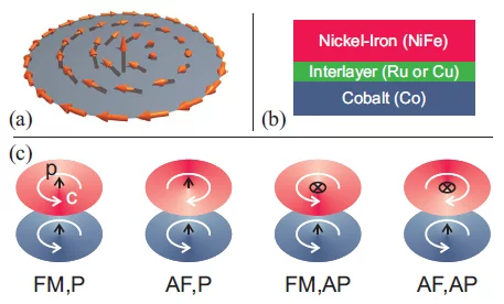
We report on the coupling of spin vortices in magnetic multilayer elements. The magnetization distribution in thin film disks consisting of two ferromagnetic layers separated by a nonmagnetic spacer is imaged layer-resolved by using x-ray microscopy. We directly observe two fundamentally different vortex coupling states, namely antiferromagnetic and ferromagnetic orientation of the flux directions. It is found that these states are predetermined for systems that involve a sufficiently strong interlayer exchange coupling, whereas for the case of a purely dipolar interaction both states are transformable into each other.
Simultaneous Surface and Bulk Imaging of Polymer Blends with X-ray Spectromicroscopy
Watts B, McNeill CR
MACROMOLECULAR RAPID COMMUNICATIONS 31, 1706-1712 (2010).
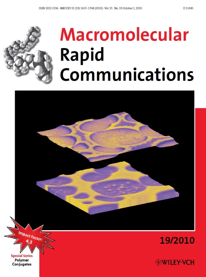
We demonstrate the utility of soft X-ray spectromicroscopy to simultaneously image the surface and bulk composition of polymer blend thin films. In addition to conventional scanning transmission X-ray microscopy that employs a scintillator and photomultiplier tube to measure the transmitted X-ray flux, channeltron detection of near-surface photoelectrons is employed to provide information of the composition of the first few nanometers of the film. Laterally phase-separated blends of two polyfluorene co-polymers are studied, with the structure of both wetting and capping layers clearly imaged. This new information provides insight into the connectivity of bulk and surface structures that is of particular relevance to the operation of such blends in optoelectronic devices.
Press Releases: PSI, Materials Views
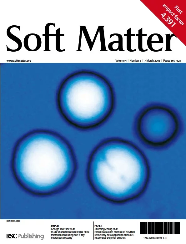
In Situ Characterization of Gas-Filled Microballoons using Soft X-ray Microspectroscopy
Tzvetkov G, Graf B, Fernandes P, Fery A, Cavalieri F, Paradossi G and Fink RH
SOFT MATTER 4, 510-514 (2008).
In this study we describe the first direct, real-space characterization of a novel type of poly(vinyl alcohol) (PVA) based microballoons in aqueous environment using scanning transmission X-ray microscopy (STXM). From the oxygen K-edge near-edge X-ray absorption fine structure (NEXAFS) spectra taken from the microballoon interiors we could unambiguously distinguish between water- and air-filled particles. We also demonstrate that STXM imaging below and above the O K-edge (520 eV and 550 eV) can provide unique information on the composition of microballoons in water. It was found that the particular microballoon system investigated here has a remarkable high stability and is able to contain gases for more than 6 months, making it well suited for biomedical applications.

