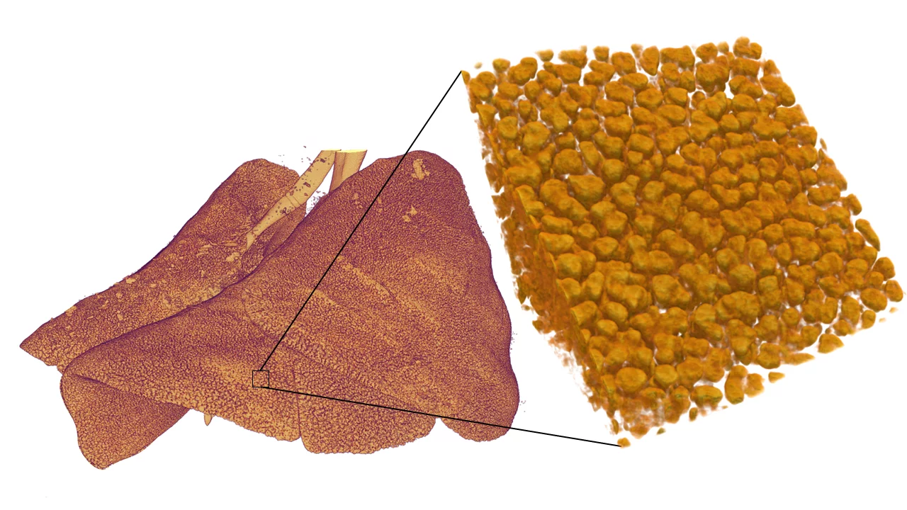
With respiratory diseases being amongst the leading causes of morbidity and mortality affecting more than 450 million people worldwide [1], X-ray computed tomography (CT) remains the gold-standard technique for detection, diagnosis, and follow-up in lung diseases. However, due to the ionizing character of X-rays, the tolerable radiation dose currently imposes the ultimate limit to the accessible resolution. In this collaborative project, we aim at improving the spatial resolution of (pre-clinical) thoracic X-ray CT beyond the physical resolution limit by using prior knowledge about the branching and microstructure of the lung learned from experimental animal models.
Fueled by the growth of computational capabilities and data availability, recent years have witnessed a surge in data-driven methods which have shown great potential in a variety of X-ray CT problems such as reconstruction [2], denoising [3] or phase retrieval [4]. In this project, we are developing data-driven super-resolution methods to recover high-resolution details from low-dose, low-resolution measurements. Our approach aims at learning a high-resolution representation of the 3D lung microstructure that can be adapted to the low-resolution measurements, incorporating the image formation process of phase-contrast thoracic CT. We hope to contribute to a deeper understanding of lung function and disease by reconstructing the entire lung at sub-resolution scale, as well as potentially find broader applications with other imaging modalities and samples where a priori knowledge about the structure exists.
This project is conducted in collaboration with the Swiss Data Science Center (SDSC Hub at PSI), the PSI Center for Scientific Computing and the Institute of Anatomy of the University of Bern.
References
- Momtazmanesh at al. “Global burden of chronic respiratory diseases and risk factors, 1990–2019: an update from the Global Burden of Disease Study 2019”, eClinicalMedicine, 2023. DOI: 10.1016/j.eclinm.2023.101936
- S. van Gogh “Hybrid tomographic reconstruction in grating interferometry breast CT: combining physics-based likelihoods with data-driven priors”, ETH Zurich. DOI: 10.3929/ethz-b-000636724
- Hendriksen at al, “Noise2Inverse: Self-Supervised Deep Convolutional Denoising for Tomography”, IEEE Transactions on Computational Imaging, 2020. DOI: 10.1109/TCI.2020.3019647
- Zhang et al, “PhaseGAN: A deep-learning phase-retrieval approach for unpaired datasets”, Optics Express, 2021. DOI: 10.1364/OE.423222
Collaborators
- Dr. Benjamín Béjar Haro, Swiss Data Science Center Hub a PSI
- Dr. Markus Janousch, PSI Center for Scientific Computing
- Alain Studer, PSI Center for Scientific Computing
- Prof. Dr. Johannes C. Schittny, Institute of Anatomy, University of Bern
Funding
PSI Research Grant
Contact
- Daniel Vera Nieto: daniel.vera-nieto@psi.ch
- Dr. Goran Lovrić: goran.lovric@psi.ch
