

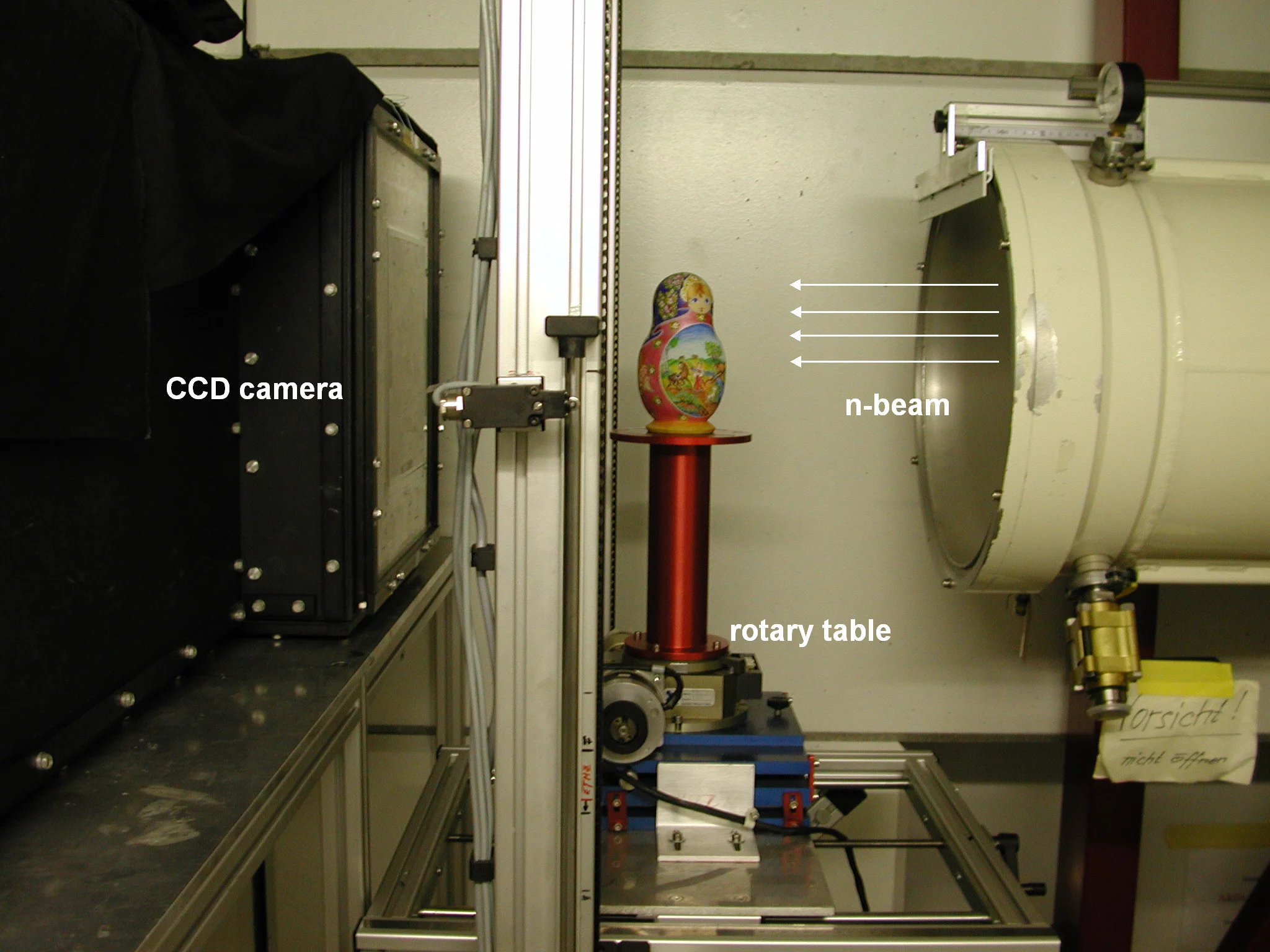
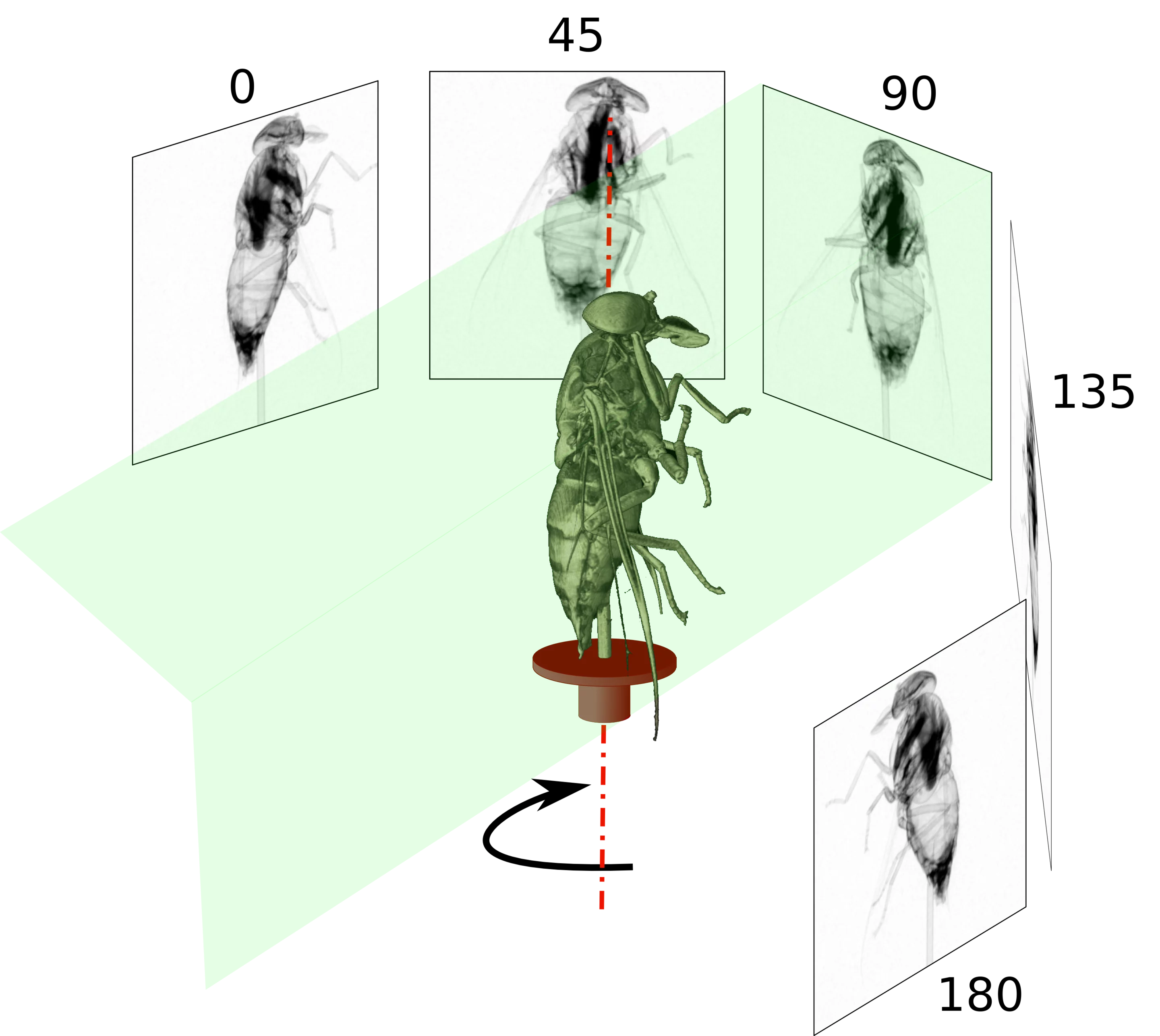
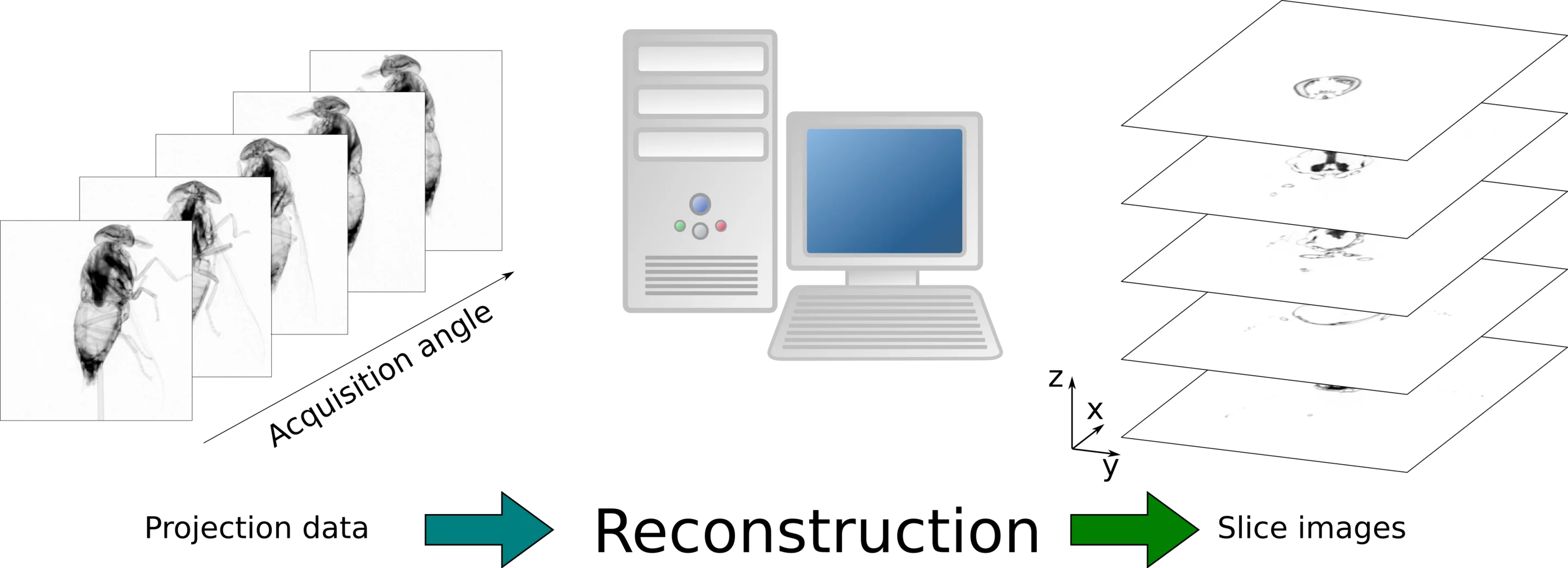
Computed tomography is a method to acquire three dimensional information about the structure inside a sample. The method applies to neutron as well as the more known X-ray imaging. It uses radiographic projection images from many views to reconstruct the distribution of materials in the sample. Mostly, the projections are acquired with equiangular steps over either 180o or 360o to cover the whole sample. Figure 9 shows an experiment setup used for neutron tomography (NT). In contrast to medical imaging the samples is rotated instead of the beamline. The projection images are acquired using a combination of a scintillator to convert the neutrons to visible light and a CCD camera. The transform of the projection data into a three dimensional image is a computationally intensive task handled by special reconstruction software. During the reconstruction process, slices perpendicular to the rotation axis are produced. When these slices are stacked in a sequence they form a three-dimensional volume image of the sample. The reconstructed volume data can be visualized using three-dimensional rendering graphics software. Using such tools, regions can be segmented based on their attenuation coefficients and geometry. This can be used to reveal details inside the sample in three dimensions.
