

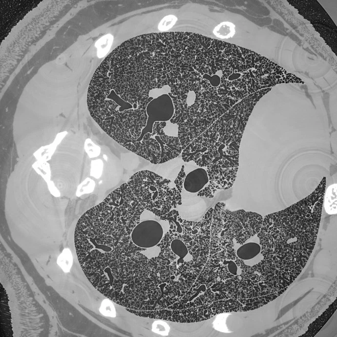
An international team of researchers, headed by the TOMCAT group and collaboration partners at the University of Bern, have used this method to acquire micrometer-scale resolution datasets on the entire lung structure of a juvenile rat in its fresh natural state within the animal’s body and without the need for any fixation, staining or other alteration that would affect the observed structure (Borisova et al, 2020, Histochem Cell Biol ). This openly accessible 1.2 TB dataset (http://link.springer.com/article/10.1007/s00418-020-01868-8) reveals the smallest structural features of the lung, the alveoli, over the whole extent of the lung.
This fast large-volume tomographic imaging method allows researchers to study the lung development in animal models with age as well as the effects of lung diseases such as asthma, chronic obstructive pulmonary disease, lung fibrosis, or pneumonia-like infections over the different stages during the course of the illness. In a next step, the regional microscopic effects and efficacy of pharmacological and other medical interventions can be investigated in detail. Furthermore, the precise knowledge of the geometry of the lung’s airways system during a particular disease pathology helps in the development of suitable pharmaceutical aerosol sprays to assure the active ingredients are deposited efficiently in the affected lung regions.
Borisova, E., Lovric, G., Miettinen, A., Fardin L, Bayat S, Larsson A, Stampanoni M, Schittny JC, Schlepütz CM. Micrometer-resolution X-ray tomographic full-volume reconstruction of an intact post-mortem juvenile rat lung. Histochem Cell Biol (2020). (http://link.springer.com/article/10.1007/s00418-020-01868-8)