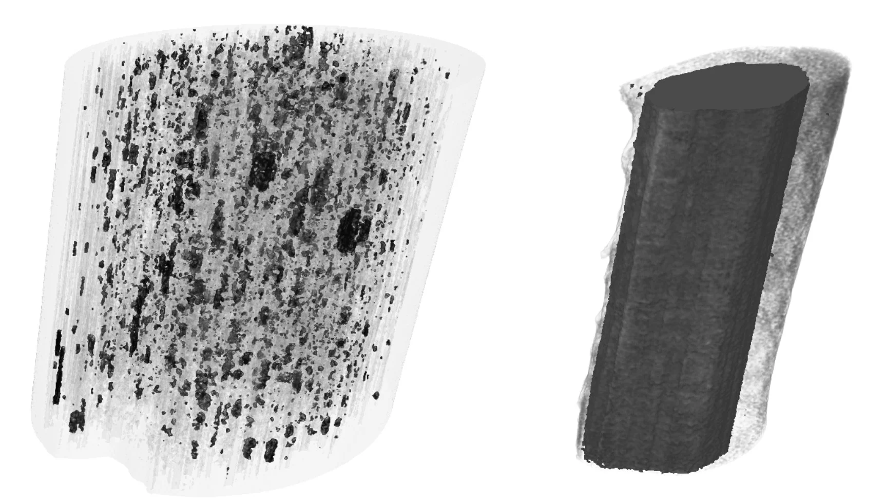
Novel carbon materials are promising candidates for light and robust low-cost materials of the future. Understanding their mechanical properties benefits from highly resolved three-dimensional (3D) maps of their porosity and density fluctuations in uninterrupted
Supplementary video
Read the full story
representativevolumes, but these are difficult to obtain with conventional imaging methods. Scientists at the Paul Scherrer Institut have now succeeded to produce in collaboration with Honda R&D in Germany highly resolved 3D density maps of entire sections of carbon fibers. The technique they used, called ptychographic computed tomography, offers unprecedented insights into the nanomorphology of these materials. Without the need of sectioning the fibers, their porosity can be visualized in 3D as can high-density carbon regions attributed to different degrees of graphitization, indicative of atomic structure differences in the material. Such imaging capabilities are expected to prove useful for the systematic study of the mechanical properties of carbon fibers, addressing a crucial point when designing and tailoring novel carbon materials.
Supplementary video
Read the full story
Reference
Characterization of carbon fibers using X-ray phase nanotomographyA. Diaz, M. Guizar-Sicairos, A. Poeppel. A. Menzel, O. Bunk
Carbon 67 (2013) 98-103, DOI:j.carbon.2013.09.066
Contact
Dr. Ana Diaz, Swiss Light SourcePaul Scherrer Institute, 5232 Villigen PSI, Switzerland
Phone: +41 56 310 5626, e-mail: ana.diaz@psi.ch
