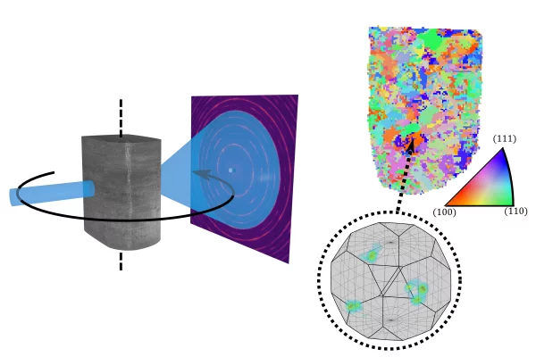

The high-penetrating power of hard x-rays along with the sensitivity to crystalline order via x-ray diffraction (XRD) makes it the one of the most important characterization-tools for crystalline materials. Thanks to the high brilliance of modern synchrotron x-ray sources, such as the Swiss Light Source at PSI, an XRD-measurement can be performed in a fraction of a second, allowing mapping of the microstructure in three dimensions from hundreds of thousands of measured diffraction patterns.
With x-ray scattering tensor tomography, materials with nanometer-sized crystallites have been mapped, giving new insight into the structure of human bone. 3D-XRD on the other hand have been used to map metals and geological materials, that contain large well-ordered crystallites. For a range of materials in-between, where the crystallites are neither small enough for tensor-tomography nor large enough for 3D-XRD, no such method has existed until now.
Such materials with micrometer-sized and deformed crystallites are not rare in real-world materials. Hardening in steel relies on generating such small-grained and deformed crystal grains and also in bio-minerals, the biological processes under which the crystallites are formed tend to create such disordered microstructures.
In this study, we demonstrate that a new approach to the data-processing, called texture-tomography, can be used bridge this gap between tensor-tomography and 3D-XRD. This new development opens the possibility to study the evolution of microstructure in-situ as a response to various processing steps and other external stimuli.