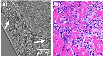
Remodelling cardiovascular diseases cause changes in size, shape or density of structural components of the heart, from whole organ to cellular level. In transplant rejection follow up, a routinely clinical procedure performed is the extraction of endomyocardial biopsies (EMBs), with consequent qualitative analysis by means of histological images. This is a time tedious process that results in tissue alteration and the assessment of a few 2D histological images for the analysis of a much larger tissue volume. Besides EMBs, full thickness samples from several regions of the heart are commonly extracted from explanted hearts and stored in the clinics for historical collection. The analysis of these samples is of prime interest for a better assessment of the reasons driving the heart towards heart failure and have the potential to give a more complete understanding.
The main goal is to propose X-ray phase contrast imaging as a fast complementary cost-effective non-destructive 3D quantitative approach for diagnosis in human EMBs and full thickness samples. Synchrotron propagation-based phase contrast imaging is used in order to identify meaningful markers at different stages of pathological remodelling and thus provide a detailed characterization of normal/abnormal tissue samples [1,2]. This information will be useful for a future implementation of the techniques in the clinics, potentially by means of lab-source based grating interferometry.
Publications
- Dejea, Hector, et al. "Microstructural analysis of cardiac endomyocardial biopsies with synchrotron radiation-based x-ray phase contrast imaging." International Conference on Functional Imaging and Modeling of the Heart. Springer, Cham, 2017.
- Dejea, Hector, et al. "Synchrotron X-ray phase contrast imaging and deep neural networks for cardiac collagen quantification in hypertensive rat model." International Conference on Functional Imaging and Modeling of the Heart. Springer, Cham, 2019.
Collaboration
- Universitat Pompeu Fabra (UPF), Barcelona, Spain
- University Hospital Centre Zagreb, Zagreb, Croatia
- IDIBAPS, Barcelona Spain
- Universitätsspital Zürich (USZ), Zürich, Switzerland
Funding
- Personalized Health and Related Technologies (PHRT) of the ETH Domain, grant #2017-33
- EU FP7 for research, technological development and demonstration under grant agreement VP2HF (no. 611823)
- Spanish Ministry of Economy and Competitiveness (gTIN2014-52923-R, the Maria de Maeztu Units of Excellence Programme MDM-2015-0502)
- FEDER
