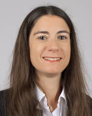
Biography
During my PhD at the INSA de Lyon (France), I studied the physical properties of materials in order to discriminate explosive to common materials (see my PhD here). Then, I spent four years in the X-ray imaging group at the European Synchrotron Radiation Facility (Grenoble, France). After a first post-doc in collaboration between the CEA-Cadarache and the ID22Ni beamline, where I gained experience in X-ray diffraction and X-ray phase contrast tomography, I was NSF Research Fellow for a Paleontology project linking the Harvard University and the ID19 beamline of ESRF. In April 2014, I joined the X-ray Tomography Group at PSI as scientific collaborator in the Phase Contrast core of the CIBM (EPFL). Since January 2016, I am Beamline Scientist at the TOMCAT Beamline. (See also my Linkedin Profile.)
Scientific Research
Specialized in X-ray imaging (micro and nano-tomography, single-distance phase-retrieval) and powder diffraction (XRD-CT), my research interest focuses on developing new imaging methods to characterize materials and understand their behaviour. Since 2014, I am in charge of the TOMCAT nanoscope. This full-field imaging setup allows getting a 3D spatial resolution of around 150 nm between 8-20 keV for a field of view of ˜70µm2. The setup can be used for absorption or phase-contrast imaging. In this case, Zernike Phase Contrast (ZPC) is exploited to enhance contrast for phase objects.
As local contact, I collaborate with international researchers on material sciences related (e.g. nuclear materials, explosives identification, cement hydration, ...) as well as palaeontology (hominid dental microstructure, fossil as rock slabs). Since 2016, I develop actively the bioimaging program at the beamline (cardiac imaging, middle-ear bones study, whole mouse brain vasculature visualisation, investigations on rodent models for neurodegenerative disease studies,...) and more particularly, I lead the Heart Imaging Project. The main goal of this international project is to quantify the cardiac macro and micro-structure using synchrotron-based X-ray phase-contrast imaging (X-PCI). We aim at providing new insights into the cardiac morphology at the micrometre scale, non-destructively, without the use of contrast agents and in 3D. With the high resolution provided by X-PCI, we can have access for instance the myofibres arrangement, vessels and trabeculation organization, and how they change with disease. In this project, we also aim to relate the structural properties of the heart and its physiological function thanks to the implementation of a modified Langendorff setup allowing us to study the heart dynamically. All those information are crucial for future clinical applications, not only to understand heart remodelling and the different patterns in cardiovascular disease but also to provide more personalized and adapted treatments. For this purpose, we are developing dedicated acquisition methodologies but also new analysis techniques based on Machine Learning algorithms.
In the context of the SLS2.0 exciting upgrade project, our team is preparing TOMCAT to push our current capabilities towards Multiscale – Multimodal – Dynamic Tomographic X-ray Microscopy with two beamlines: S-TOMCAT and I-TOMCAT.
Selected Publications
For an extensive overview we kindly refer you to our publication repository DORA.
Visualizing plating-induced cracking in lithium-anode solid-electrolyte cells
Ning Z., Spencer Jolly D., Li G., De Meyere R., Pu D. S. , Chen Y., Kasemchainan J., Ihli J., Gong C., Liu B., Melvin L.R. D., Bonnin A., Magdysyuk O., Adamson P., Hartley O. G., Monroe W. C. , James Marrow T. and Bruce P. G., Nat. Mater. (2021).
Additive manufacturing of silica aerogels
Zhao, S., Siqueira, G., Drdova, S., Norris, D., Ubert, C., Bonnin, A., Galmarini S., Ganobjak M., Pan Z., Brunner S., Nyström G., Wang J., Koebel MM and Malfait, W. J. (2020). Nature, 584(7821), 387-392.
Liquid flow and control without solid walls
Dunne P, Adachi T, Dev AA, Sorrenti A, Giacchetti L, Bonnin A, Bourdon C, Mangin PH, Coey JMD, Doudin B & Hermans TM (2020) Nature, 581(7806), 58-62
Synchrotron radiation imaging revealing the sub-micron structure of the auditory Ossicles
Anschuetz L, Demattè M, Pica A, Wimmer W, Caversaccio M and Bonnin A, Hearing Research. 2019; 383: 107806
Comprehensive analysis of animal models of cardiovascular disease using multiscale X-ray phase contrast tomography
Dejea H, Garcia-Canadilla P, Cook AC, Guasch E, Zamora M, Crispi F, Stampanoni M, Bijnens B and Bonnin A, Scientific Reports. 2019; 9(1): 6996
NRStitcher: non-rigid stitching of terapixel-scale volumetric images
Miettinen A, Oikonomidis IV, Bonnin A. and Stampanoni M, Bioinformatics. 2019: btz423
X-ray Fourier ptychography,
Wakonig K, Diaz A, Bonnin A, Stampanoni M, Bergamaschi A, Ihli J, Guizar-Sicairos M and Menzel A, Science Advances. 2019; 5(2): eaav0282
An up-to-date publication list can be found in my publons account.
