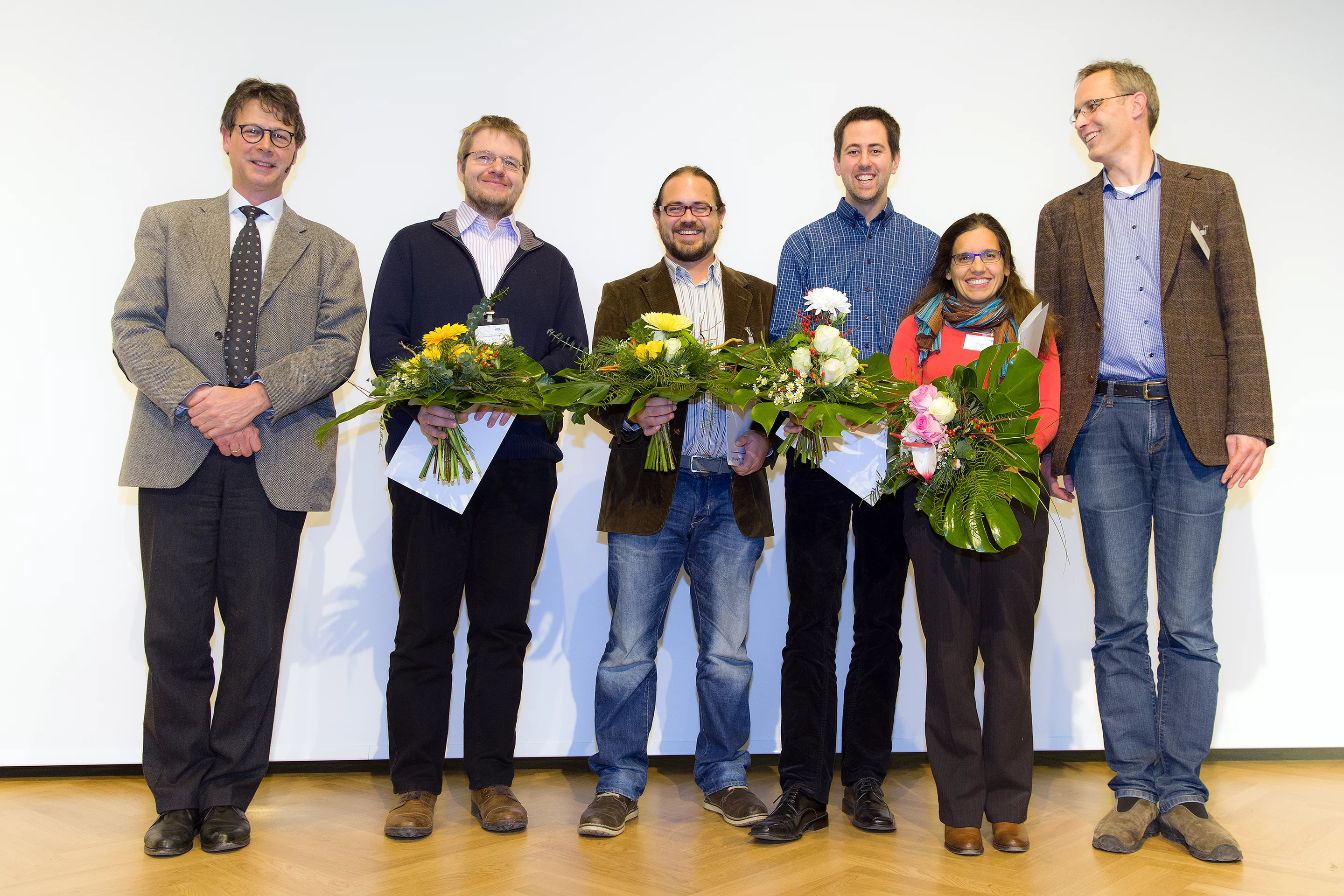
The 2014 Innovation Award on Synchrotron Radiation was bestowed to researchers Ana Diaz, Manuel Guizar-Sicairos, Mirko Holler, and Jörg Raabe from the Paul Scherrer Institut, Switzerland, for their contributions to method and instrumentation development, which have set new standards in high-resolution 3D hard X-ray microscopy.
The team utilizes and develops a promising X-ray microscopy technique called ptychography, which is a scanning variant of coherent diffractive imaging (CDI). This technique exploits coherence properties of synchrotron X-ray sources and, using computing algorithms in lieu of lenses, obtains high-resolution quantitative 3D images of electron density. Recently they imaged a nanoporous glass structure of 6 microns diameter at an isotropic 3D resolution of 16 nm – a resolution record in X-ray tomography of non-isolated specimens. This resolution achievement was made possible by continuously improving both instrumentation and analytical algorithms over the past years. A 3D high-precision scanning stage was developed and commissioned, with positions measured and stabilized via dedicated laser interferometry that incorporates a rotational degree of freedom for tomography. Simultaneously, improvements in post-processing and alignment of projections proved crucial, and algorithmic developments help better to exploit the measured signals.
The measurements are currently performed at higher than 6.2 keV photon energies, such that thick specimens can be accurately characterized. Even though ptychography is a scanning technique, a tremendous speed above of 25,000 resolution elements per second has been demonstrated recently. The 3D resolution and quantitative electron density contrast opens up new fields of application for hard X-ray 3D microscopy. These first demonstrations paved the way to studying sample systems with unprecedented detail, ranging from biological tissue over cement to catalytic particles.
Developments continue. Now that the positioning concept is proven, Mirko Holler endeavors to finalize OMNY, the tomography nano cryo stage, with a vacuum environment and cryogenic temperatures to preserve the structural integrity of radiation-sensitive samples. For this a new set-up has been designed and machined in collaboration with the AMI department. The set-up is currently being assembled and first measurements will start soon. Due to the provided phase and absorption contrast, at cryogenic conditions even weakly scattering objects like biological networks or cellular tissue could be measured without staining or fixation.
The highly prestigious award is granted yearly by the Association of Friends of the Helmholtz-Centre Berlin for “an excellent achievement which has contributed significantly to the further development of techniques, methods or uses of synchrotron radiation.” The team received the award in Berlin in a ceremony on December 4, 2014.
Links
- The Association of Friends of the Helmholtz-Centre Berlin
- Innovation Award on Synchrotron Radiation
- Fast scanning coherent X-ray imaging using Eiger
- Sixteen nanometers in 3D
Related publications
An instrument for 3D x-ray nano-imagingM. Holler, J. Raabe, A. Diaz, M. Guizar-Sicairos, C. Quitmann, A. Menzel, and O. Bunk
Rev. Sci. Instrum. 83, 073703 (2012) DOI:10.1063/1.4737624
X-ray ptychographic computed tomography at 16 nm isotropic 3D resolution
M. Holler, A. Diaz, M. Guizar-Sicairos, P. Karvinen, E. Färm, E. Härkönen, M. Ritala, A. Menzel, J. Raabe & O. Bunk
Scientific Reports 4, 3857 (2014) DOI:10.1038/srep03857
High-throughput ptychography using Eiger: scanning X-ray nano-imaging of extended regions
M. Guizar-Sicairos, I. Johnson, A. Diaz, M. Holler, P. Karvinen, H.-C. Stadler, R. Dinapoli, O. Bunk, and A. Menzel
Optics Express, 22, 14859-14870 (2014) DOI:10.1364/OE.22.014859
Contact
Dr. Mirko Holler, Swiss Light SourcePaul Scherrer Institut, 5232 Villigen PSI, Switzerland
Phone: +41 56 310 3613, e-mail: mirko.holler@psi.ch