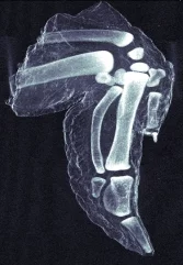
A type of X-ray imaging that shows detail otherwise lost, and which is compatible with conventional radiography instrumentation is now feasible, reports a study published online in Nature Materials. This technique offers unprecedented resolution for several applications, including medical imaging, security screening and industrial non-destructive testing.
Dark-field imaging is commonly used in visible light microscopy, and it enables details to be resolved that are otherwise smeared out in the direct reflection mode or bright field. The quality of the dark-field image depends on the intensity of the light scattered by the object. With X-rays, however it has always been difficult to have a high enough signal-to-noise ratio, and therefore the use of X-ray dark-field imaging has usually been restricted to very-high-intensity light sources, such as synchrotrons.
In a collaborating research team led by Franz Pfeiffer (Laboratory for Synchrotron Radiation) and Christian David (Laboratory for Micro-and Nanotechnology) has shown that by using an appropriate arrangement of certain optical components it is possible to obtain a high signal-to-noise ratio even with conventional X-ray tubes, as they demonstrate by revealing the very fine structure of bones in a chicken wing.
Read full article
Facility: SLS
Hard-X-ray dark-field imaging using a grating interferometer, Franz Pfeiffer* , Martin Bech, Oliver Bunk, Philipp Kraft, Eric F. Eikenberry, Christian Brönnimann, Christian Grünzweig and Christian David, Nature Materials , 174801 (2007) AOP: http://dx.doi.org/10.1038/nmat2096 (20.01.2008)
Hard-X-ray dark-field imaging using a grating interferometer, Franz Pfeiffer* , Martin Bech, Oliver Bunk, Philipp Kraft, Eric F. Eikenberry, Christian Brönnimann, Christian Grünzweig and Christian David, Nature Materials , 174801 (2007) AOP: http://dx.doi.org/10.1038/nmat2096 (20.01.2008)
