General Layout
An overview of the beamline hutches is given below. The superbending magnet photon source and the front-end are to the left, inside the machine tunnel. A small sample-preparation and machine lab is situated at the rear of the beamline.
Source
The following table shows the main properties of the X-ray source for SuperXAS.
| Ring energy | eE | 2.4 | GeV |
|---|---|---|---|
| Ring current | eI | 400 | mA |
| Magnetic field | B | 2.9 | T |
| Critical Energy | Ec | 11.1 | keV |
| Electron beam size | σX | 46.3 | µm |
| Electron beam size | σY | ~11 | µm |
| Electron beam emittance | εX | 5.63 | nm rad |
| Electron beam emittance | εY | 0.007 | nm rad |
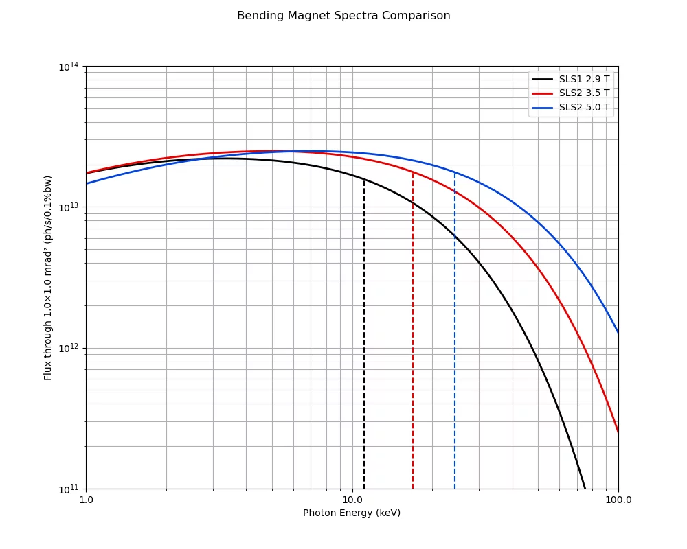
Spectras of the superbending magnets are shown in the image below. The current superbending magnet with 2.9 T will be replaced during the SLS Upgrade to a superbending magnet with a variable field strength of 3.5 T or 5 T (changeable during machine shutdowns).
Optics
The optical components of the SuperXAS beamline, their functions and position from the source are listed here.
| Beamline component | Function | Distance from the source [mm] |
|---|---|---|
| Graphite filter unit | Beam attentuation | 5970 |
| White beam slits | Definition of beam divergence | 6364 |
| Collimating mirror M1 | Vertical collimation | 7744 |
| QEXAFS Monochromator MO1 | Fast scanning, monochromatic beam | 12980 |
| Bremsstrahlungs stopper | Block white- and pink-beam | 13635 |
| Monochromatic slits | Beam definition (block scattering beam) | 14145 |
| Beam profile monitor | Monitoring beam profile | 14525 |
| Focusing mirror M2 | Bendable toroid, focusing the beam | 15580 |
| Beam profile monitor | Monitoring beam profile | 17276 |
| Harmonic rejection mirror | Rejection of high order harmonics | ~22600 |
| Main focus spot | Main position for sample | ~23600 |
X-ray mirrors
The high stability mirror system is designed to be very robust, reliable and to allow easy access for installation and maintenance. A very important design aspect of the mirrors systems is the complete mechanical separation between the optics and the vacuum chamber. This aims to avoid as much as possible vibrations propagation to the mirrors. A massive granite block directly supports the mirrors holders/benders that are mechanically isolated from the vacuum chamber by means edge-welded bellows.
The mirrors can be remotely adjusted in five independent degrees of freedom where the rotations (pitch, roll and yaw) are realized by means of software pseudo motors. The two (02) UHV compatible translation stages installed in the vacuum chamber with a UHV compatible stepper motor for each stage allow horizontal translation of the mirror. Thus, coating can be changed between Pt and Rh (plus Si coating for M1).
Vertical translations are performed by means of three (03) vertical jacks that also perform the pitch and roll rotations.
| Property | Collimating mirror (M1) | Focusing mirror (M2) |
|---|---|---|
| Mirror substrate | Monocrystalline silicon | |
| Direction of Reflection | Upwards | Downwards |
| Shape | Flat, cylindrically bent to tangential cylinder | Sagittal cylinder, cylindrically bent to torus |
| Tangential bending radius | 4.5 km to flat (>40 km) | 3.0 km to flat (>40 km) |
| Sagittal bending radius | Flat |
20 mm (Pt coated) 30 mm (Rh coated) |
| Optical useful length | 1000 mm | |
| Optical useful width | 3 x 20 mm (Si, Rh, Pt) | 2x 34 mm (Rh, Pt) |
| Sagittal slope error | < 10 µrad RMS | < 20 µrad RMS |
| Tangential slope error | < 1.5 µrad RMS | < 2.5 µrad RMS |
| Micro roughness | < 3 Å RMS | |
| Coating |
> 60 nm Rh, Pt Cr underlayer |
|
| Cooling | Max power load ~100 W, clamped side watercooling | No cooling |
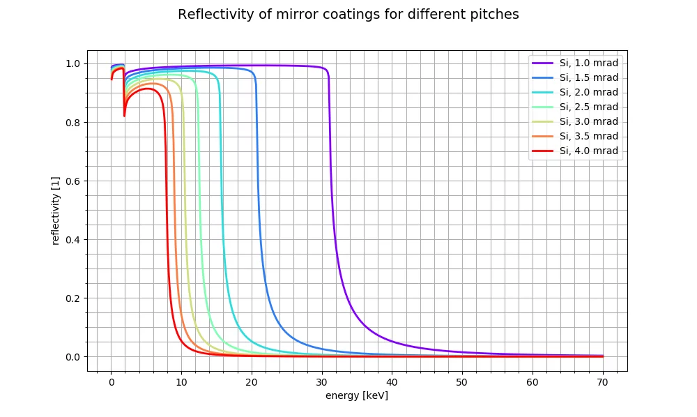
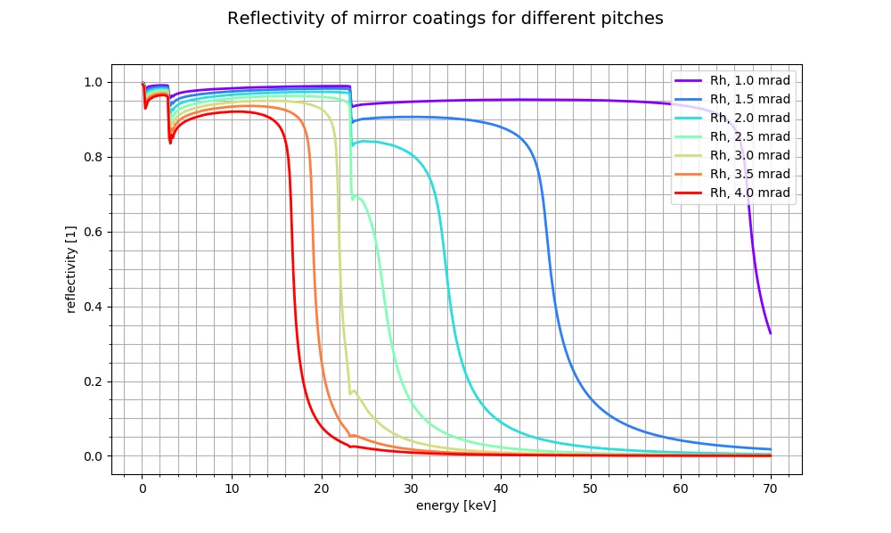
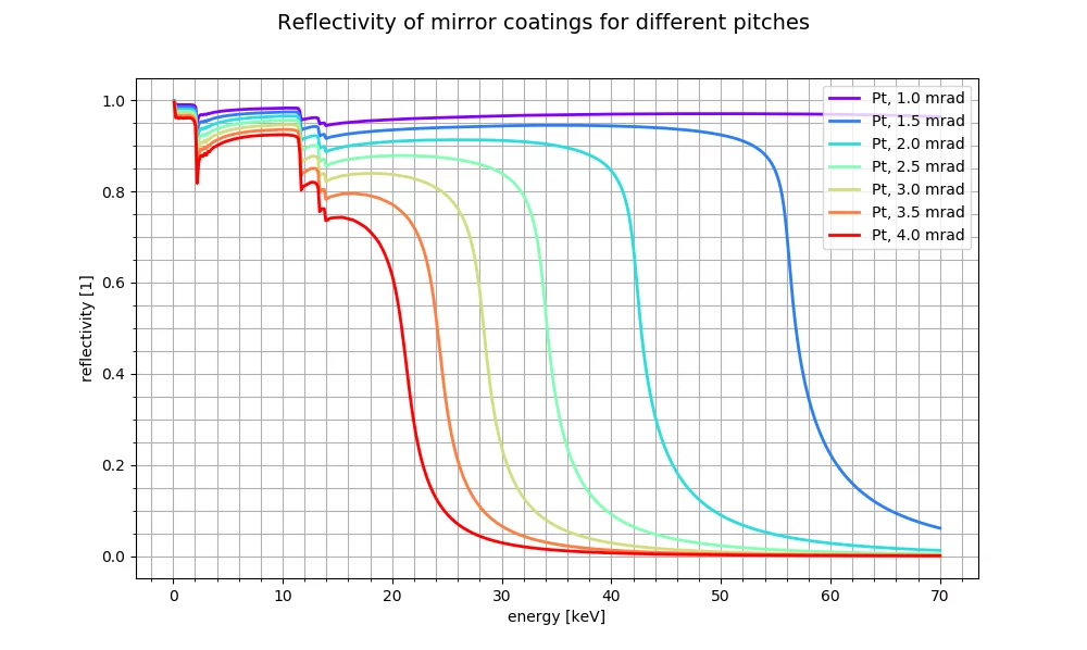
The reflectivities of different mirror pitches are shown below for the mirror surfaces used at SuperXAS. Click on the image to enlarge.
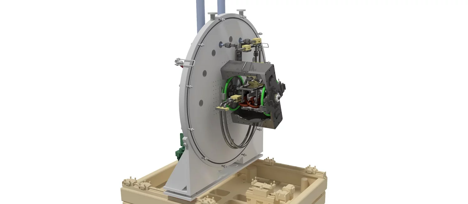
Quick EXAFS monochromator (QEXAFS)
The QEXAFS monochromator consists of two channel cut crystals attached to a direct-drive torque motor. The system facilitates scan speeds for a specific energy range with up to 20 spectra per second (full EXAFS). Additionally, for low concentration samples, the monochromator can be operated in step-scanning mode by using the goniometer. The operational energy range can be selected by changing between the Si(111) and the Si(311) crystal and is in the range of 4.5 to 25 keV for the Si(111) and 9 keV to 35 keV for the Si(311). The crystals are cryo-cooled with LN2.
The system is a further development of the monochromator developed by Uni Wuppertal in Germany:
-
Müller O, Nachtegaal M, Just J, Lützenkirchen-Hecht D, Frahm R
Quick-EXAFS setup at the SuperXAS beamline for in situ X-ray absorption spectroscopy with 10ms time resolution
Journal of Synchrotron Radiation. 2016; 23: 260-266. https://doi.org/10.1107/S1600577515018007
DORA PSI


