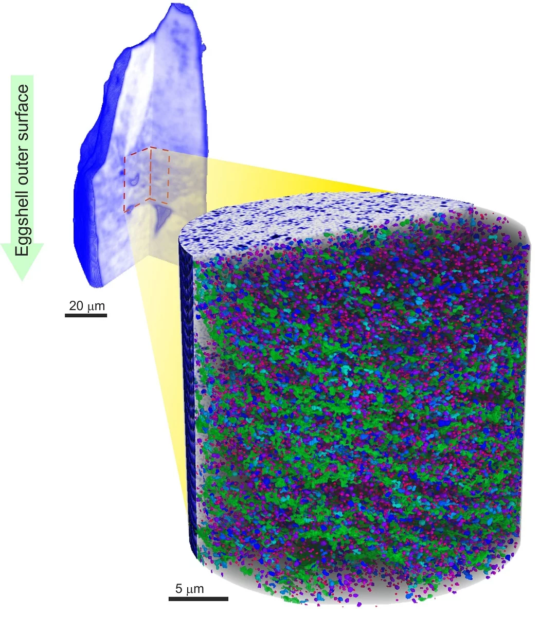
In nature and materials science macroscopic properties are often optimized by hierarchical structures in which relevant organization occurs at many different length scales, spanning a range covering nanometers, microns, and millimeters. The study of such structures benefits from imaging in 3D at different zoom levels to understand them in detail. However upon zooming into a region of interest within the object an inherent problem in tomography arises, termed sometimes the “interior problem,” and by which image quality is reduced and a direct interpretation of the contrast in the image is forgone. The problem is solvable with prior information about the sample but this is not always available. Researchers at PSI have developed a technique that combines measurements at different resolution levels, effectively a 3D overview and a detailed interior inspection, which disambiguates the interior reconstruction and preserves image quality and quantitative interpretation. In collaboration with researchers of the institute AMOLF in the Netherlands and ETH Zurich in Switzerland they showcase their technique by studying the porous structure within a section of an avian eggshell. The section itself has a diameter of around 60 micrometers, about the size of a human hair. The detailed measurements of the interior of the sample allowed the researchers to quantify the ordering and distribution of an intricate network of pores within the shell.
The need and ability to zoom in to get higher resolution is ubiquitous in imaging and allows one to access information about progressively finer details. In our everyday experience we physically approach an interesting placard to take a better look, or use our camera zoom to enlarge our subjects. Laboratory microscopes are usually equipped with different microscope objectives that provide a range of different magnifications. In this way a low resolution overview is available to study large-scale features and the overall environment and higher magnifications can be used for more detailed studies of interesting regions.
X-ray tomography provides 3D images with invaluable information on 3D structure and composition in the life and materials sciences, in essence providing views of virtual slices within the sample without the need to dissect or destroy it. Tomography for medical applications allows one to reach resolutions below a millimeter. However, dedicated instruments can also obtain high-resolution 3D images at resolutions below one micrometer (one thousand of a millimeter). A technique named ptychographic X-ray computed tomography, developed at the Swiss Light Source, utilizes the high brilliance of synchrotron light and can reach imaging below 20 nanometers, which is 20 millionths of a millimeter. This technique also provides accurate mass density characterization of materials. Such unique combination is currently used to study natural and synthetic materials at these minute length scales.
The problem of interior tomography
Although in many cases the hardware to image very different length scales exists, for tomography it is somewhat more difficult to zoom into a region of interest compared to 2D imaging. Tomography requires images of the object of interest from many different angles, and these views are later combined by a tomography reconstruction algorithm to obtain a 3D image. Zooming in, in this case means to image a small volume of interest within the full object, which is referred to as interior tomography. The so-called “interior problem” arises with the realization that the tomographic reconstruction of an interior region is not well-posed because not all the sample is measured at all the necessary orientations. The measurement is then incomplete and cannot be accurately reconstructed. Although a great deal of information of shape, size, and orientation can be obtained from interior reconstructions, artefacts do appear and the image values can no longer be interpreted quantitatively.
There has been a significant amount of work in addressing the “interior problem” for tomography, and it has been repeatedly shown that very little additional information about the sample is sufficient to remove artifacts and recover quantitative contrast. In previous work such additional information is either used during reconstruction using advanced algorithms or by adjusting the final images to comply with already known values at some sample locations. This information is however not always available.
Combining multiresolution tomograms to get a clear view
Researchers at the Swiss Light Source developed an alternative approach that allows nanoscale quantitative density studies with ptychographic X-ray tomography on interior regions of a sample without using any assumptions or prior information. They make up for the incomplete measurement by using a low-resolution 3D overview of the sample. By combining the interior high-resolution measurements with this faster and significantly lower-resolution overview they disambiguate the interior reconstruction and overcome limitations and artefacts arising from tomography of an internal region of interest.
In their work published in Optica, they studied avian eggshell as a model for a bio-nanoporous structure. Eggshell possesses a complex hierarchical structure and provides robust protection while serving as a calcium reservoir for the developing embryo. The overview provided a 3D image with about one micron resolution where gas exchange pore channels, necessary for oxygenating the embryo, were visualized. It also hinted at the presence of layers of nanoscale vesicles, but they were too small to be seen in this coarse overview. High-resolution interior measurements revealed this nanoscale porosity with detail thus far only observed by electron microscopy in 2D, in contrast to the 3D information retrieved in this study. The quantitative-contrast reconstructions enabled by this technique allowed for reliable segmentation for detailed analysis of orientation of vesicles and vesicle layers.
Nanoporous materials are ubiquitous in biology and offer valuable insight into growth and development, their structure achieves a remarkable structural robustness while minimizing the used material. Study of these materials in multiple scales sheds light on the structural basis for the development of strong and light synthetic materials.
Besides multiscale imaging applications there is marked interest in the solution of the “interior problem” for tomography. Reliable interior imaging is also an active topic of research for medical diagnostics as it localizes the radiation dose on the region of interest. Here we experimentally demonstrate that quantitative interior tomography is feasible by combination with a low-resolution overview, the latter taking close to 100 times less radiation dose. The demonstrated approach has wide applicability at a wide gamut of length scales and even beyond X-ray contrast.
PSI release
From inside an eggshell.Multimedia
Supplementary video showing the overview and interior tomogram as well as the porosity analysis.Reference
Quantitative interior x-ray nanotomography by a hybrid imaging techniqueManuel Guizar-Sicairos, Jaap J. Boon, Kevin Mader, Ana Diaz, Andreas Menzel, and Oliver Bunk
Optica 2, 259--266 (2015) DOI:10.1364/OPTICA.2.000259
Contact
Dr. Manuel Guizar-Sicairos, Swiss Light SourcePaul Scherrer Institut, 5232 Villigen PSI, Switzerland
Phone: +41 56 310 3409, e-mail: manuel.guizar-sicairos@psi.ch