
Tiny voids inside eggshells supply the materials that stimulate and control the shell’s growth. Using a novel imaging technique, researchers from the Paul Scherrer Institute (PSI), ETH Zurich and the Dutch FOM Institute AMOLF have succeeded in imaging these voids in 3D for the first time. In doing so, they lift an old limitation of tomographic images and hope that one day medicine will also benefit from their method.
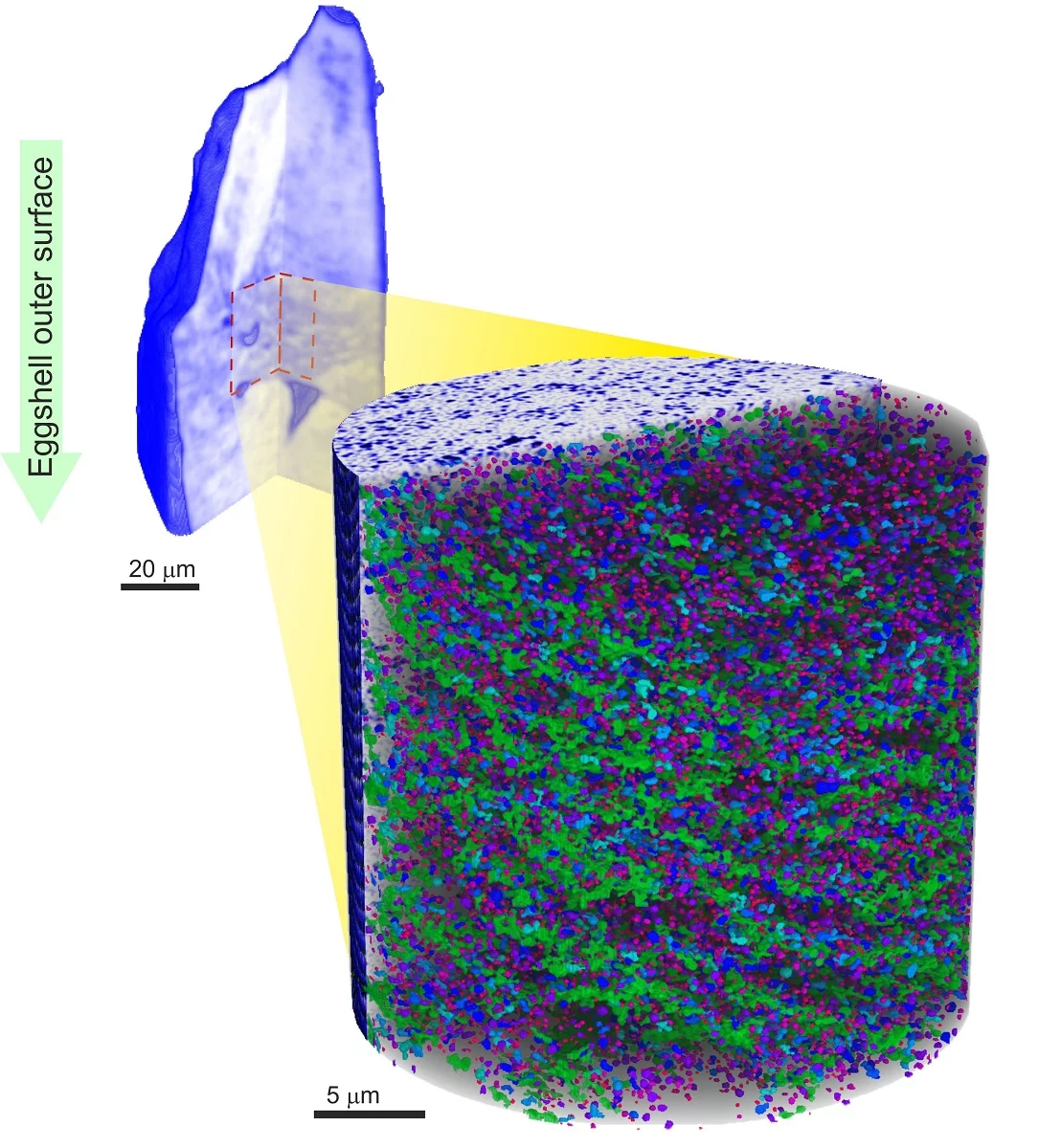
Researchers from the Paul Scherrer Institute (PSI), ETH Zurich and FOM Institute AMOLF in the Netherlands have developed a method that enables them to produce extremely detailed 3D images of sections of an object with the aid of x-ray light. They used the technique to render visible a network of nanometre-sized holes in the shell of a chicken’s egg, for which only two-dimensional pictures were previously available. The x-ray images were produced at the beamline cSAXS at PSI’s Swiss Light Source (SLS). The eggshell does not just protect chicks against the harsh outside world. It also stores calcium, which the chicks need to grow, and it lets in air so the embryos can breathe. The structure of the eggshell forms a matrix made of biocalcite mineral (calcium carbonate), through which the outside air passes via micrometre-sized pores. Even smaller than these pores are the voids that deliver those substances that control the shell’s growth.
Solution for an old imaging problem
Thanks to a novel imaging technique, a team of scientists headed by Manuel Guizar-Sicairos from PSI have now succeeded in rendering the nanometre-sized vesicles in the eggshell visible in three dimensions. Previously, it was only possible to image these bubbles with the aid of electron microscopes, which provide two-dimensional images. The researchers have now produced an extremely detailed 3D image of the vesicle network in a small section of an eggshell – about the size of a human hair – using highly brilliant x-ray light from the SLS. Not only were they able to display this network with nanometre resolution, but also obtain quantitative information about the density of the surrounding material. Determining the density confirmed that this material was, as expected, calcite. To obtain these results, the authors of the study were able to tackle an old imaging problem: it was previously impossible to only image one section of an object in 3D in so much detail, without compromising the resolution and quantitative information content.
Sharpness thanks to blurred overview
The problem is rooted in how these tomographic images are produced. As the object to be depicted needs to be illuminated with x-ray light from many different directions, it is turned on an axis perpendicular to the x-ray light. A two-dimensional detector placed behind the object records the x-ray light that the object has altered. The researchers can then reconstruct a 3D image of the object on the computer from these 2D images, each of which forms a pattern of light and dark areas. If the researchers now want to produce a 3D image of a section from inside this object – i.e. virtually zoom in on the section in question – the image quality suffers. The problem stems from the fact that only parts of the section’s surroundings are exposed when it is illuminated. This loss of information makes the subsequent image reconstruction on the computer difficult as the reconstruction programs need this information on the surroundings to obtain quantitative information about the section.
By implication, this means that practically the entire object needs to be strongly illuminated in order to image the section in sufficient detail. And this in turn means a lot of time and an undesirably high level of radiation exposure for the object to be depicted. It is as if a person’s whole head had to be strongly illuminated, even though you only wanted a sharp image of the nose. The question that tomography experts have been grappling with for some time: how can the section be imaged as quickly, in as much detail and with as low a radiation dosage for its surroundings as possible? Or to use the same comparison, how can the nose be depicted sharply without strongly illuminating the whole head? Guizar-Sicairos’s team has now found the answer: it is enough to combine a strongly illuminated image of the section with a less detailed image of the entire object. In other words, the nose is strongly exposed and the rest of the head weakly, which is sufficient to produce a sharp 3D image of the nose. The less sharp overview image can be taken quickly and still provides sufficient information to reconstruct the section in high resolution. Overall, this results in a lower radiation dose around the region of interest and a smaller amount of time needed for the image.
Medical applications conceivable
The new technique could be used to image sections of an object in high resolution more quickly, without burdening the object’s surroundings unnecessarily strongly with radiation. This could also make the method interesting for medical applications. Computer images of part of an organ would be conceivable, for instance, without having to irradiate the patient’s whole body intensely. The radiation could therefore be focused on the part of an organ that one wants to study, largely sparing the surrounding tissue. In terms of hardware, no major obstacles should stand in the way of the new technique as it does not depend on x-ray light from a large-scale facility like the SLS. “With suitable adjustments, it would also work with the x-ray machines used in hospitals,” says Guizar-Sicairos.
Text: Paul Scherrer Institute/Leonid Leiva
Additional information
Coherent X-ray scattering groupContact
Dr. Manuel Guizar-Sicairos, Coherent X-rax scattering group, Paul Scherrer Institute,Telephone: +41 56 310 34 09, E-mail: manuel.guizar-sicairos@psi.ch
Original Publication
Quantitative interior x-ray nanotomography by a hybrid imaging techniqueManuel Guizar-Sicairos, Jaap J. Boon, Kevin Mader, Ana Diaz, Andreas Menzel, and Oliver Bunk
Optica 2, 259--266 (2015)
DOI:10.1364/OPTICA.2.000259