

The TOMCAT beamline at the Swiss Light Source specializes in rapid high-resolution 3-dimensional tomographic microscopy measurements with a strong focus on biomedical imaging. The team has recently developed a technique to aquire large volume datasets with high resolution in a very short time.
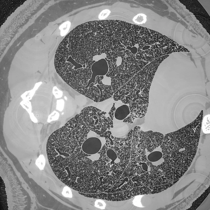
Micrometer-resolution X-ray tomographic full-volume reconstruction of an intact post-mortem juvenile rat lung
An international team of researchers, headed by the TOMCAT group and collaboration partners at the University of Bern, have used this method to acquire micrometer-scale resolution datasets on the entire lung structure of a juvenile rat in its fresh natural state within the animal’s body and without the need for any fixation, staining or other alteration that would affect the observed structure (Borisova et al, 2020, Histochem Cell Biol ). This openly accessible 1.2 TB dataset (http://link.springer.com/article/10.1007/s00418-020-01868-8(link is external)) reveals the smallest structural features of the lung, the alveoli, over the whole extent of the lung.
For further information, please click here.
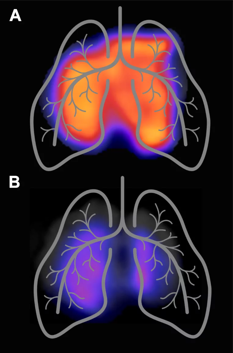
Imaging Lung Inflammation using PET: The Use of 18F-AzaFol and beyond
The Center for Radiopharmaceutical Sciences (CRS) focuses on the development of new radiopharmaceuticals for positron emission tomography (PET) and other applications in nuclear medicine. Researchers at CRS have previously developed an 18F-based PET tracer (18F-AzaFol) for targeting the folate receptor, which is expressed on tumor cells, but also on activated macrophages involved in inflammatory processes. In a recent clinical study, 18F-AzaFol was investigated regarding its in vivo properties for folate-receptor targeting and assessed with respect to its safety profile in patients (NCT03242993).
For further information, please click here.