Fast and accurate data collection for macromolecular crystallography using the JUNGFRAU detector
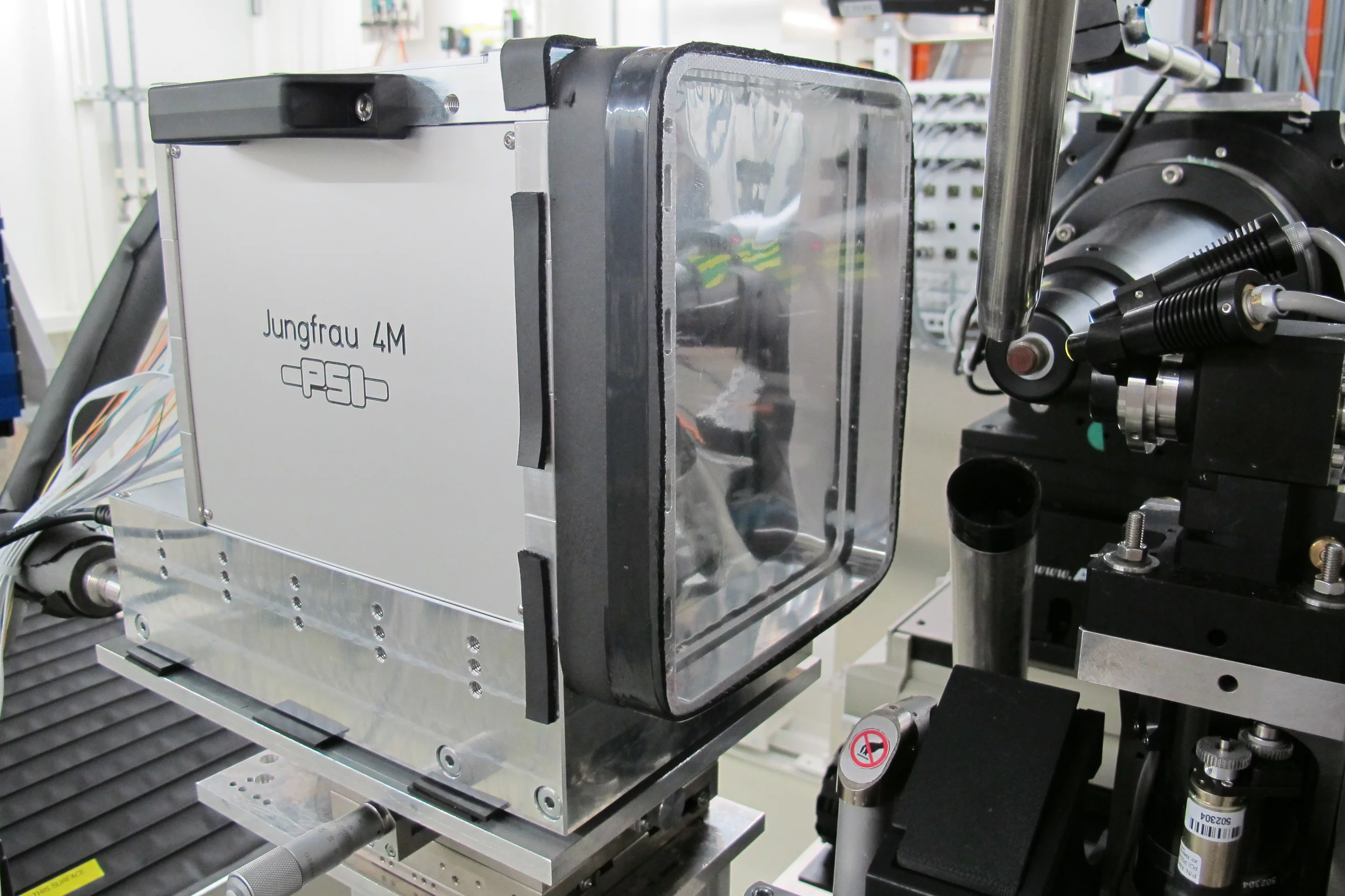
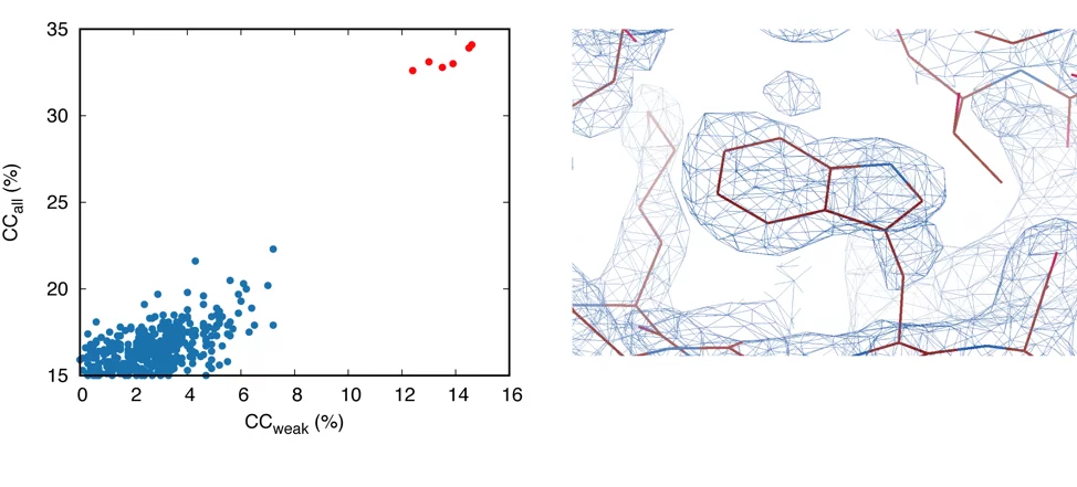
The JUNGFRAU detector represents the latest generation of pixel-array detector from the Paul Scherrer Institut. It uses direct detection charge integrating technology with dynamic gain switching to achieve a high dynamic range while maintaining a low-noise performance. Compared to previous generation X-ray detectors, JUNGFRAU improves the accuracy of crystallographic measurements in two respects. First it removes count-rate limitation inaccuracies for high photon counts, allowing the full flux of the next generation light sources to be used for faster measurements in Macromolecular Crystallography (MX). At the same time JUNGFRAU improves the accuracy of long wavelength data collection for de novo structure determination using native-SAD phasing, due to uniform pixel sensitivity. We have demonstrated both effects by showing that a 600 millisecond exposure of a thaumatin crystal with full X-ray flux (6 keV) is sufficient to capture the weak anomalous signal accurately enough for experimental phasing (Figure 1). The results are reported in the October 2018 issue of Nature Methods [1,2].
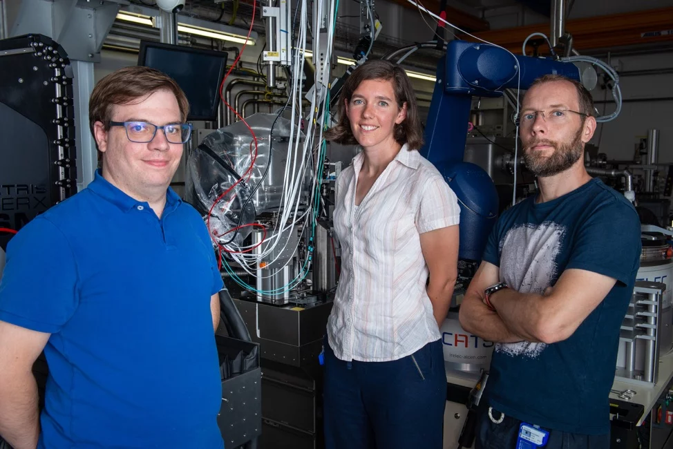
While JUNGFRAU was designed for SwissFEL, the free electron laser now in operation at PSI, this work demonstrates the advantage that JUNGFRAU technology can also bring to synchrotrons. However, in addition to the evident benefits in terms of data accuracy, the JUNGFRAU brings associated challenges when applied to synchrotron applications. The anticipated data rates are unprecedented, with a data stream of 20 Gbit/s produced per million detector pixels. Near to real-time data handling has been achieved with a JUNGFRAU 4 Mpixel detector running at 1.1 kHz in our latest tests (Figure 2, 3). Active developments are currently underway at PSI to construct a JUNGFRAU with more than 10 million pixels for MX applications at the SLS and to further increase the frame rate to 2.4 kHz.
Contact
Dr. Bernd Schmitt, Swiss Light SourcePaul Scherrer Institut, 5232 Villigen PSI, Switzerland
Phone: +41 56 310 2314, e-mail: bernd.schmitt@psi.ch
Dr. Meitian Wang, Swiss Light Source
Paul Scherrer Institut, 5232 Villigen PSI, Switzerland
Phone: +41 56 310 4175, e-mail: meitian.wang@psi.ch
Publications
1. Fast and accurate data collection for macromolecular crystallography using the JUNGFRAU detector.Leonarski F., Redford S., Mozzanica A., Lopez-Cuenca C., Panepucci E., Nass K., Ozerov D., Vera L., Olieric V., Buntschu D., Schneider R., Tinti G., Froejdh E., Diederichs K., Bunk O., Schmitt B., Wang M.
Nat. Methods 15, 799–804 (2018)
DOI: 10.1038/s41592-018-0143-7
2. A detector for the sources.
Chapman H. N.
Nat. Methods 15, 774–775 (2018)
DOI: 10.1038/s41592-018-0150-8