Time is essential in many experiments, in particular, if processes are studied. We may for example want to know how liquids move through porous media or quantify the shape of a solidification front.
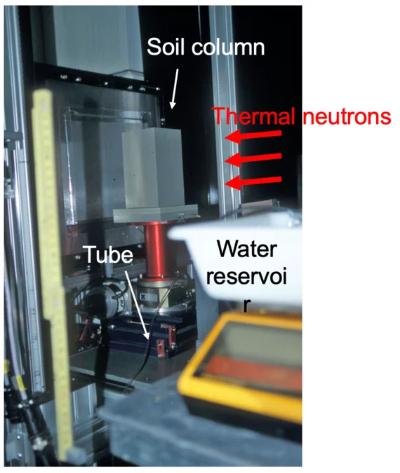
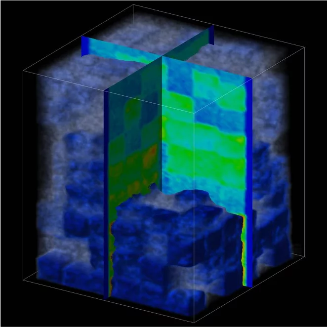
Early on-the-fly experiments were performed to understand wetting and drainage processes in heterogeneous porous structures made of sands with different grain size distributions [1].
In [2], Zarebanadkouki et al. observe and model the water flow in the rhizosphere and in particular within the roots. This study showed that there are different pathways for the water in the days than during the night. Another example using on-the-fly tomography can be found in [3] where the front of cadmium contaminated water in sandstone is studied.
These are processes on the time scale of neutron imaging and have often been studied using time series of radiographs. The problem is that changes caused by the processes in many cases must be studied in three dimensions. The three-dimensional images are obtained using tomography. However, tomography requires that the sample doesn’t change significantly for the entire duration of a scan. Otherwise, there will be motion artifacts in the reconstructed images.
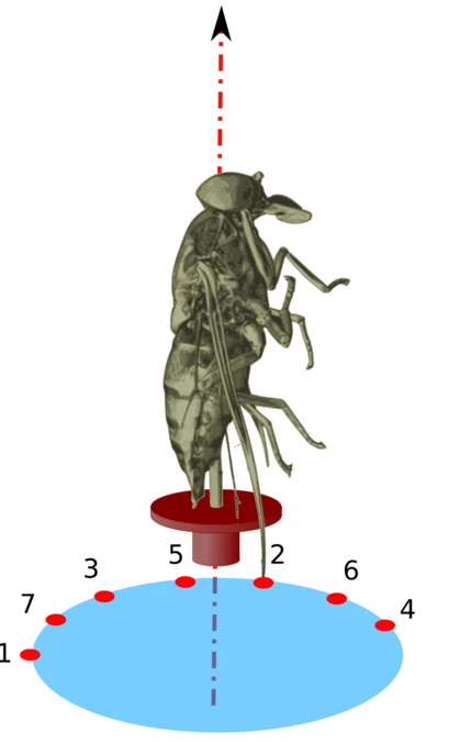
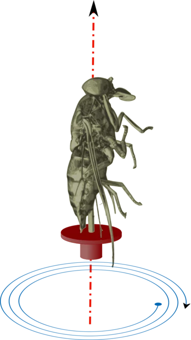
There are different ways to reduce motion artifacts that appear when the sample changes over time. The choice depends on how fast the process is, the needed pixel size to detect the spatial changes, and what noise levels can be accepted. The options are shown in the figures below:
- Quasi-steady state where the process is stopped at different states to make the tomography.
- Slow processes can be scanned using an acquisition scheme based on the golden ratio [1]. This scheme has the advantage that the number of volumes can be chosen after the scan. The technique was developed for experiments like [6].
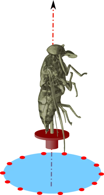
- The fastest processes can be studied using the on-the-fly method [1,5]. With this technique, the sample is rotated with constant speed while images are continuously acquired. Neutron tomography scans are normally performed stepwise with small angular increments.
The challenge of the latter two techniques is to household with the available neutrons to obtain the image quality needed to study the intended phenomenon.
Publications
[1] Kaestner A, Hassanein R, Vontobel P, Lehmann P, Schaap J, Lehmann E and Flühler H 2007 Mapping the 3D water dynamics in heterogeneous sands using thermal neutrons Chemical Engineering Journal 130
[2] Zarebanadkouki M, Trtik P, Hayat F, Carminati A, and Kaestner A 2019 Root water uptake and its pathways across the root: quantification at the cellular scale Scientific Reports 9
[3] Cordonnier B, Pluymakers A, Tengattini A, Marti S, Kaestner A, Fusseis F and Renard F 2019 Neutron Imaging of Cadmium Sorption and Transport in Porous Rocks FRONTIERS IN EARTH SCIENCE 7
[4] Kaestner A, Münch B, Trtlk P and Butler L 2011 Spatiotemporal computed tomography of dynamic processes Optical Engineering 50
[5] Zarebanadkouki M, Carminati A, Kaestner A, Mannes D, Morgano M, Peetermans S, Lehmann E and Trtik P 2015 On-the-fly Neutron Tomography of Water Transport into Lupine Roots Physics Procedia vol 69
[6] Trtik P, Münch B, Weiss W J, Kaestner A, Jerjen I, Josic L, Lehmann E and Lura P 2011 Release of internal curing water from lightweight aggregates in cement paste investigated by neutron and X-ray tomography Nuclear Instruments and Methods in Physics Research, Section A: Accelerators, Spectrometers, Detectors and Associated Equipment 651
[7] Wyrzykowski M, Ghourchian S, Münch B, Griffa M, Kaestner A and Lura P 2021 Plastic shrinkage of mortars cured with a paraffin-based compound – Bimodal neutron/X-ray tomography study Cement and Concrete Research 140 106289
Contact
- Dr. Anders Kaestner, anders.kaestner@psi.ch, +41 56 310 42 86
- Dr. Pavel Trtik, pavel.trtik@psi.ch, +41 56 310 55 79
