
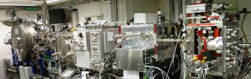
General beamline description and specifications
The PHOENIX (PHotons for the Exploration of Nature by Imaging and XAFS) beamline is dedicated to X-ray Absorption (micro-) Spectroscopy (XAS) and imaging in the soft and the rarely served tender X-ray energy range of 0.3 to 8 keV. The beamline covers important absorption edges of low-Z elements, in particular the K-edges of N-Fe and the L –edges of Ca-Er, providing unique research opportunities for material science, biology, energy research, environmental science, chemistry, catalysis or cultural heritage.The beamline has two branchlines. The PHOENIX I branch uses a double crystal monochromator and covers energies from 1-8 keV, the soft x-ray branchline PHOENIX II invokes a planar grating monochromator and covers the range from 0.3-2 keV.
The source for both branchlines is an Apple-II undulator, which is time shared with the X-Treme beamline.
All experiments are performed in a vacuum chamber (ca. 10-5 -10-6 mbar), and both branchlines offer ample opportunities for complex user supplied in situ experiments.
| Energy range | 0.8 - 8.0 keV PHOENIX I 0.4 - 2.0 keV PHOENIX II |
|---|---|
| Photon source | elliptical APPLE II undulator for linear and elliptical polarization |
| Flux (3keV) | 1 x 10¹¹ ph/s/0.1%BW/400 mA |
| Energy resolution | 1 x 10⁻⁴ |
| Focused spot size | 2.5 µm x 2.5 µm |
| Sample environments | - solid samples, pellets - measurement in vacuum or helium - possible heating to 450 C, cooling to 90K - titration cells, flow-through liquid cells - liquid microjet (in collaboration) |
| Fluorescence detectors | 4 element and 1 element silicon drift diode (SDD) energy dispersive detectors |
| von Hamos spectrometer |
Crystal radius 7 cm, segmented crystal. Energy range for standard von Hamos geometry: Si111 : 2200 (2030) -3100 eV Si 220 : 3330 (3250 )-5060 eV Energies in brackets represent lowest energies reachable upon request using small samples. |
Access to the PHOENIX beamline
To access the PHOENIX beamline, please consult the user information user information available on this website.
The Phoenix Group is part of the Laboratory for Synchrotron Radiation and Femtochemistry (LSF).
Measurement opportunities at PHOENIX
X-ray absorption spectroscopy (XAS) and chemical imaging
PHOENIX offers opportunities for high quality spectroscopy (i.e. EXAFS) and imaging of light elements, with the possibility of studying samples with low concentration. The beamline is equipped with two different end-stations providing state-of-the-art equipment to conduct X-ray micro-spectroscopic measurements (µ-XAS and µ-XRF) using fluorescence, transmission and total electron yield as detection methods. Chemical imaging of bulk samples can be done with a spatial resolution in the micrometer range using x-ray scanning microscopy.
X-ray emission spectroscopy (XES)
A von Hamos spectrometer for tender x-rays is available for emission spectroscopy. Using two crystals, the instruments range for routine operation is Si111 : 2200 (2030)-3100 eV and for Si 220 : 3330 (3250)-5060 eV. The energies given in brackets, are the lowest possible energies (by request only). The energy resolution is in the order of 0.5 eV or better. The spectrometer is completely integrated into the routine microscopy setup, and hence can be coupled to imaging applications. For further information about the spectrometer, please contact Thomas Huthwelker.
Time-resolved Studies
Furthermore, in a collaborative effort with Dr. G. Smolentsev, novel pump and probe schemes (ms to ps time resolution) for tender X-rays are currently under development and this experiment is opened for first experienced users for pilot experiments. Duetto and IDOL lasers can be used for photoexcitation.
Sample environments
Standard solid samples can be measured in vacuum or helium atmosphere. Heating to 450 C and cooling to 90 K is possible. The beamline offers different sample environments including but not limited to liquid micro-jet, liquid flow cell and in situ titration cells. We also provide a special chamber, which allows simple integration of user provided in situ experiments. Upon request dedicated sample environments can be developed within scientific collaboration.
Scientific opportunities at the PHOENIX beamline
Research at PHOENIX covers various fields, such as environmental science, catalysis, energy research, electrochemistry, biology, cultural heritage and characterization of novel materials. Here we highlight some of the fields. For a complete list of publications see PHOENIX publications.
Electrochemistry
An emerging field for tender x-rays is electrochemistry, as low-Z elements gain increasing importance. Using PHOENIX, researchers have addressed questions related to the chemistry of sulphur-based batteries (Gorlin et al., 2015), phosphorous containing batteries (Schmidt et al., 2018) and sodium based batteries (Kulka et al.,2020). Using soft X-rays, the complex interplay between O, Ni, Co in a battery using both K and L-edges have been explored (Redel et al., 2020).
Environmental science and biology
Using micro spectroscopy, questions in environmental science, soil chemistry, biology and nuclear waste management can be addressed: For example, the chemical interplay of roots with the soil environment has been studied by van Veelen et al. (2019). Vogel et al. (2020) addressed the impact of fertilizers on the chemical state of nitrogen in soil using soft X-ray. Fichtner et al. (2018) studied the state and distribution of sulfur in shells, Lange et al. (2018) used micro-spectroscopy to explore the distribution of strontium in bivalve shells. As example for biolological applications, Czapla-Masztafiak et al. (2015) compared the distribution of sulfur and phosphorous species healthy and cancerous tissue. The physio-chemical processes which cause severe concrete degradation was studied by Geng et al., 2020).
Studies on aqueous solution Structure
As XAS reflects the local environment around the probed atom, it is uniquely suited to determine the coordination of ions in solution. At PHOENIX we offer a liquid microjet system, and various flow cells for such studies. Examples for such research include investigations on the deformation of the sulfate ion in solution (Pin et al., 2015), the formation of ion paring in carbonate solutions (Henzler et al., 2018), the study of ionic distances in sodium chloride solution (Galib et al., 2017), Duignan et al. (2019), and the impact of coordination geometry of aluminum in solids and liquids on multielectron excitations (Fulton et al., (2015)
Catalysis
The tender x-ray range is of particular interest to problems in catalysis and energy research. For example, the L edges of transition metals can be covered and the formation and role of palladium carbides was studied (Tew et al., 2012; Tew et al., 2011). Another important element, aluminium, is frequently studied at the beamline. One examples is its use as dopant in catalysts (Bowker et al., 2020). Aluminosilicates (Zeolites) are important catalysts, but the location of Al in the framework is still under debate, which was explored by (Vjunov et al., 2014), who used EXAFS to locate Al atoms on certain T-sites in zeolites, and explored the role of water in the zeolite framework (Vjunov et al., 2015). Other studies address the mechanism of sulfur poisoning on Ru catalysts (Koenig et al., 2014), or the role of phosphorous in metal-organic frameworks. (Morel et al., 2015).
Crystallization and nucleation
Using a liquid microjet system and other in situ experiments, the role of ion pairing in supersaturated carbonate solutions (Henzler et al. 2018, Highlight), has been investigated by using XAS taken from a liquid microjet system in combination with theoretical modelling. XAS has also been used to study the growth and crystallization pathways of carbonates (Xto et al., 2019a; Xto et al., 2019b; Xto et al., 2019c), and the role of water in the carbonate system (Du et al., 2020, Du et al., 2019).
Cultural heritage
The beamline is suited for cultural heritage studies. Examples include studies of the discoloring of famous paintings (Cato et al., 2016), or the colour of ancient carpet hair (Meyer et al., 2016).
Opportunities for medical and biological research at the PHOENIX beamline
The PHOENIX beamline offers imaging on a micrometer scale and X-ray spectroscopy to analyze the chemical composition of a sample. Using the tender X-ray range, PHOENIX is well suited to perform X-ray micro spectroscopy on elements (O, N, F, Na, Mg, Al, Si, P, Cl, K, Ca, .., Fe), which are relevant to biological systems. This technique can be used to decipher the chemical role (oxidation state, type of chemical bonding, speciation, etc. with micrometer spatial resolution) of these elements in a biological system. For example, the chemical structure of Sulfur and Phosphorous has been studied in cancerous tissues. (e.g. Czapla-Mastiafiak, 2015).
With the current COVID-19 health crisis, we invite in particular research proposals tackling these problems. Currently, there is an open rapid access call for research projects on COVID-19: (https://www.psi.ch/en/sls/scientific-highlights/priority-access-call-for-work-on-combating-covid-19)
For possible projects at the PHOENIX beamline, please contact Thomas Huthwelker.