This branch delivers X-ray photons in the energy range of 0.8 - 8.0 keV. A monochromatized beam is delivered using one of the 4 crystal pairs mounted in the monochromator (Be, KTP, InSb and Si). Two sets of focusing mirrors (in Kirkpatrick-Baez configuration) provide a focal spot size of about 2.5 µm x 2.5 µm in two different vacuum chambers. The incoming beam intensity (I0) is monitored by a Ni covered thin polyethylene foil by measuring its drain current. Absorption measurements can be done either in transmission, with total electron yield (TEY), or in fluorescence modes (TFY). The X-ray fluorescence is detected either by a single-element or a 4-element Si drift diode array, which are sensitive to trace elements as low as a few ppms. The micro-focused beam allows the mapping of the elemental distribution in heterogeneous samples, by saving the full XRF spectra in each pixel.
First sample vacuum chamber in the beam path (standard chamber) can adapt five different stages to hold the sample and position it into the beam. The chamber is large enough (Fig. 1) to adapt in-situ setups for moderately cooling or heating the samples in vacuum, or in closed cells.
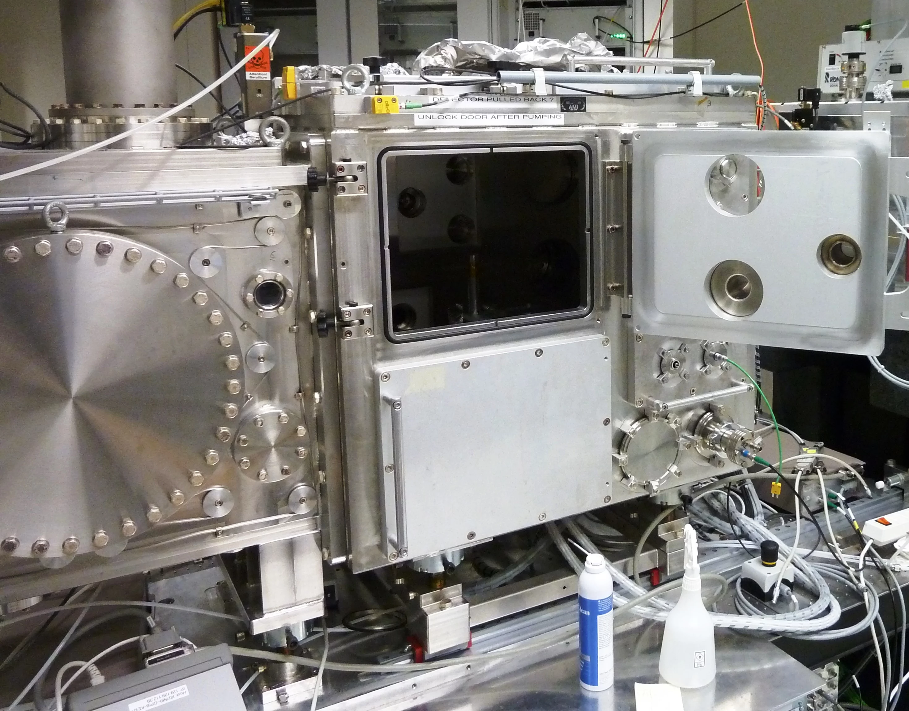
Figure 1. View of the standard chamber with focused beam size of 2.5x2.5 microns.
The standard sample holder is shown in Figure 2 for solid samples fixed on Cu plate. The available area is 40x24.5 mm. The samples can be embedded into In foil or fixed with carbon tape onto the Cu plate for a good TEY signal.
The standard sample holder is shown in Figure 2 for solid samples fixed on Cu plate. The available area is 40x24.5 mm. The samples can be embedded into In foil or fixed with carbon tape onto the Cu plate for a good TEY signal.
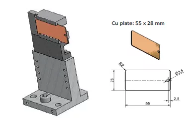
Figure 2. Schematic of the standard (solid) sample holder.
The top view of the measurement setup placed in focus position is shown in Fig. 3. The sample manipulator has 4 linear stages and 1 rotation stage for sample alignment and scanning. The standard measurements are done under 45 deg incidence with respect to the incoming beam. The online microscope which is placed at 0 deg with respect to the surface normal can be used for sample visualization with a resolution of few microns.
The top view of the measurement setup placed in focus position is shown in Fig. 3. The sample manipulator has 4 linear stages and 1 rotation stage for sample alignment and scanning. The standard measurements are done under 45 deg incidence with respect to the incoming beam. The online microscope which is placed at 0 deg with respect to the surface normal can be used for sample visualization with a resolution of few microns.
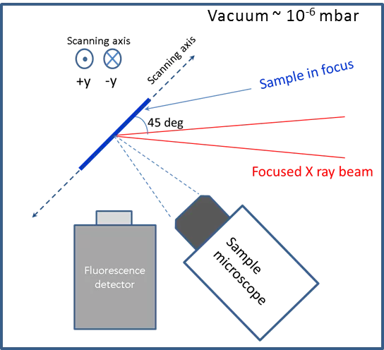
Figure 3. Top view of the measurement setup with the sample positioned in the 45 deg. geometry and in the focal planes of X-rays and on-line microscope.
A second vacuum chamber (chemistry chamber) placed in the beam path of the PHOENIX I branch is shown in Figure 4. The purpose of this smaller vacuum chamber is to host dirty experiments involving liquids or aggressive gases. The X-ray beam can be focused by a second focusing unit down to 5x5 microns.
A second vacuum chamber (chemistry chamber) placed in the beam path of the PHOENIX I branch is shown in Figure 4. The purpose of this smaller vacuum chamber is to host dirty experiments involving liquids or aggressive gases. The X-ray beam can be focused by a second focusing unit down to 5x5 microns.
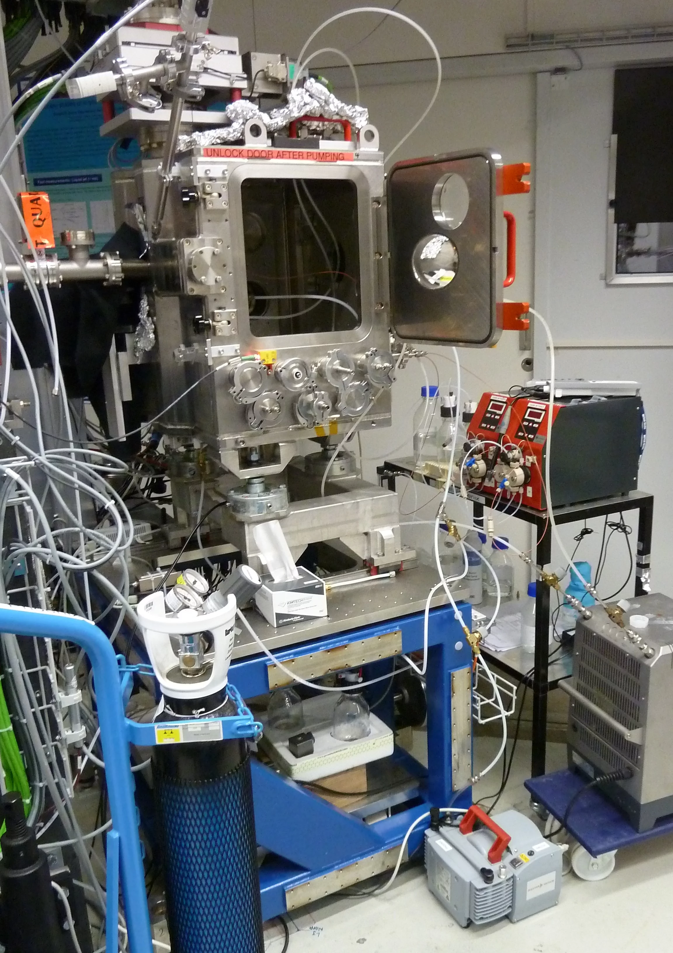
Figure 4. View of the chemistry chamber attached to PHOENIX I branch with focused beam size of 5x5 microns.
The sample manipulator is mounted from the top down, in order to protect the motors against possible spills. The available area on the Cu plate is 50x24.5 mm. The manipulator has 3 linear motors and 1 optional rotation motor for the sample alignment and scanning. The standard measurements are done similarly, under 45 deg incidence with respect to the incoming beam, and an identical online microscope is placed normal to the sample surface as in the standard chamber.
The sample manipulator is mounted from the top down, in order to protect the motors against possible spills. The available area on the Cu plate is 50x24.5 mm. The manipulator has 3 linear motors and 1 optional rotation motor for the sample alignment and scanning. The standard measurements are done similarly, under 45 deg incidence with respect to the incoming beam, and an identical online microscope is placed normal to the sample surface as in the standard chamber.
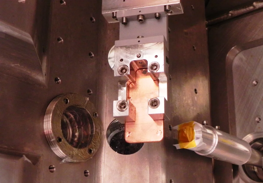
Figure 5. View of the sample holder attached to the top manipulator in the chemistry chamber.
Various liquid cells are available upon request and in collaboration for in-situ studies of chemical reactions. Figure 6 shows the drawings of the titration (5 ml volume) and flow-through liquid cells (0.1 ml volume).
Various liquid cells are available upon request and in collaboration for in-situ studies of chemical reactions. Figure 6 shows the drawings of the titration (5 ml volume) and flow-through liquid cells (0.1 ml volume).
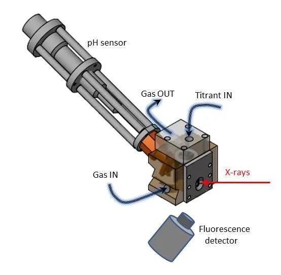
Figure 6. Schematic of the liquid titration cell (total volume 5 ml).
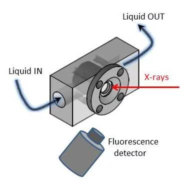
Figure 7. Schematic of the flow-through cell (total volume 0.1 ml).
Liquid jet experiments have been extensively used to understand fast chemical reactions (~ ms time scale) by mixing solutions at a throughput of 2-5 ml/min. Figure 7 shows a 100 micron diameter liquid jet placed in front of 1-element Si detector.
Liquid jet experiments have been extensively used to understand fast chemical reactions (~ ms time scale) by mixing solutions at a throughput of 2-5 ml/min. Figure 7 shows a 100 micron diameter liquid jet placed in front of 1-element Si detector.
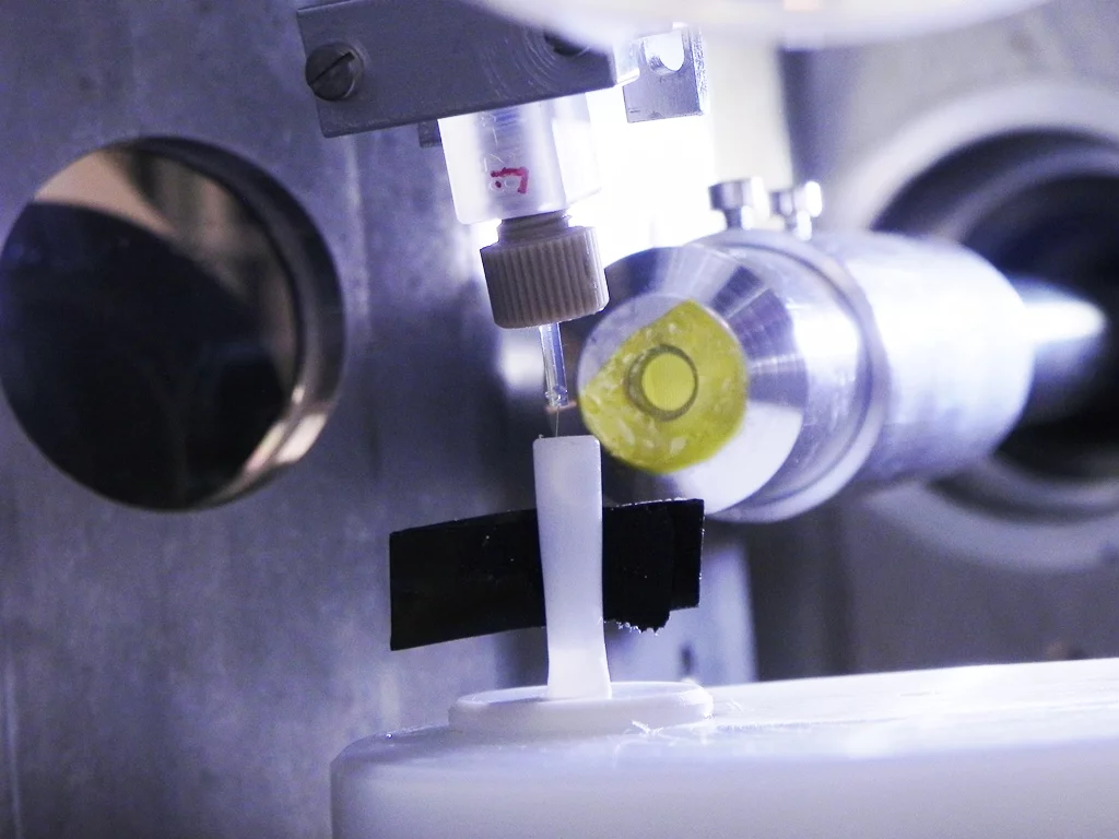
Figure 8. View of the 100 micron diameter liquid jet (flow rate 1 ml/min).
