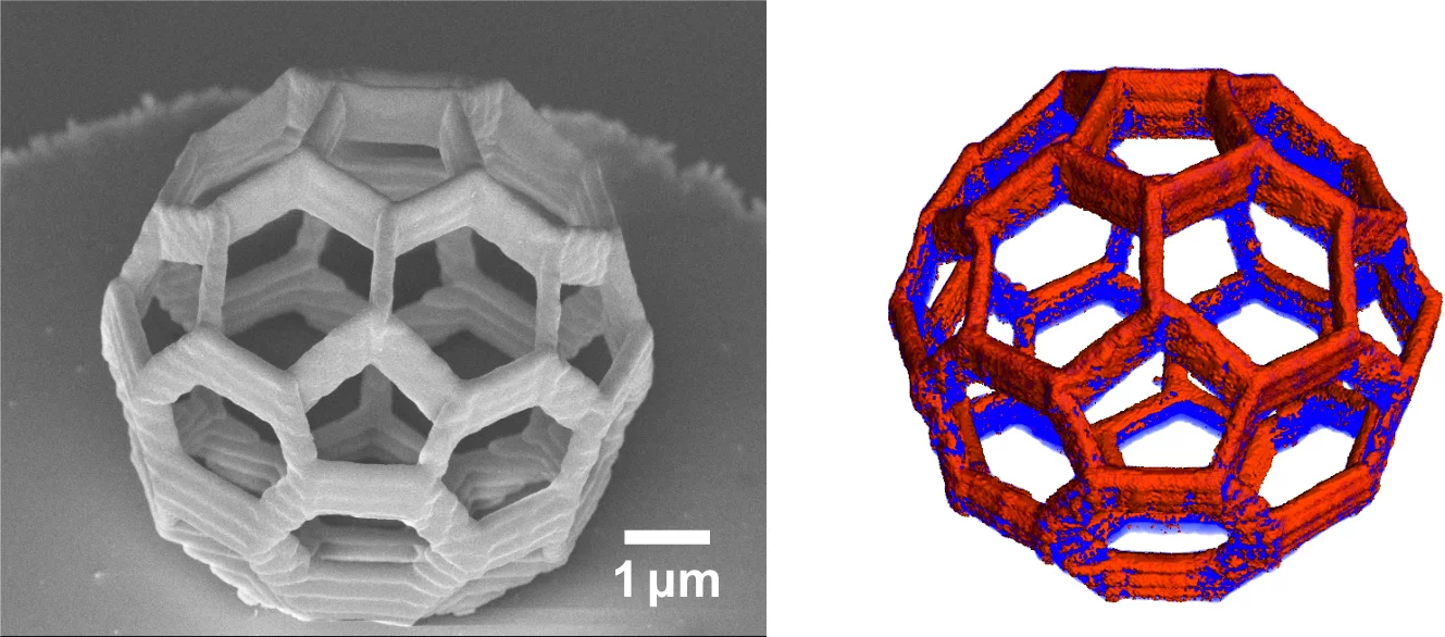
Recent advances in fabrication techniques to create mesoscopic 3D structures have led to significant developments in a variety of fields including biology, photonics, and magnetism. Further progress in these areas benefits from their full quantitative and structural characterization. We present resonant ptychographic tomography, combining quantitative hard x-ray phase imaging and resonant elastic scattering to achieve ab initio element-specific 3D characterization of a cobalt-coated artificial buckyball polymer scaffold at the nanoscale. By performing ptychographic x-ray tomography at and far from the Co K edge, we are able to locate and quantify the Co layer in our sample to a 3D spatial resolution of 25 nm. With a quantitative determination of the electron density we can determine that the Co layer is oxidized, which is confirmed with microfluorescence experiments.
PSI Media Release
Nanometres in 3DOriginal Publication
Element-Specific X-Ray Phase Tomography of 3D Structures at the NanoscaleC. Donnelly, M. Guizar-Sicairos, V. Scagnoli, M. Holler, T. Huthwelker, A. Menzel, I. Vartiainen, E. Müller, E. Kirk, S. Gliga, J. Raabe and L. J. Heyderman
Phys. Rev. Lett. 114, 115501 - Published 16 March 2015, DOI: 10.1103/PhysRevLett.114.115501
Contact
Dr. Manuel Guizar-Sicairos, Swiss Light SourcePaul Scherrer Institut, 5232 Villigen PSI, Switzerland
Phone: +41 56 310 3409, e-mail: manuel.guizar-sicairos@psi.ch
Dr. Joerg Raabe, Swiss Light Source
Paul Scherrer Institut, 5232 Villigen PSI, Switzerland
Phone: +41 56 310 5193, e-mail: joerg.raabe@psi.ch
Prof. Laura Heyderman
Laboratory for Mesoscopic Systems, Department of Materials, ETH Zurich
Laboratory for Micro- and Nanotechnology, Paul Scherrer Institute
5232 Villigen PSI; Switzerland
Phone: +41 56 310 2613, e-mail: laura.heyderman@psi.ch
