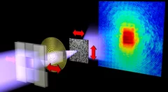
A schematic of the setup employed for the experimental demonstration. X rays are focused and scatter off a test sample that can be displaced laterally with nanometer precision. The diffraction pattern produced by the scattered X rays is collected by a detector. The sample is reconstructed on a computer from the diffraction data (see other images).
X-rays allow an inside look at structures that cannot be imaged using visible light. They are used to investigate nanoscale structures of objects as varied as single cells or magnetic storage media. Yet, high-resolution images impose extreme constraints on both the X ray microscope and the samples under investigation. Researchers at the Technische Universität München, Germany, and the Paul Scherrer Institut in Villigen, Switzerland, now showed how to relax these conditions without loss of image quality. They further showed how to image objects featuring fast fluctuations, such as the rapid switching events that determine the life time of data storage in magnetic materials. They demonstrated their method with an experiment at the Swiss synchrotron SLS and with computer simulations. The results have been published in the science journal Nature.
Read the full story
Read the full story
Original publication
Reconstructing state mixtures from diffraction measurementsPierre Thibault, Andreas Menzel, Nature, 7. February 2013
DOI: 10.1038/nature11806
Contact
Dr. Andreas Menzel; Labor für Makromoleküle und BioimagingPaul Scherrer Institut, 5232 Villigen PSI, Switzerland
Phone: +41 56 310 3711, e-mail: andreas.menzel@psi.ch [German, English]
Dr. Pierre Thibault; Physikdepartment
Technische Universität München, 85748 Garching, Germany
Phone: +49 (0)89 289 14397, e-mail: pierre.thibault@tum.de [French, English]