
X-rays allow an inside look at structures that cannot be imaged using visible light. They are used to investigate nanoscale structures of objects as varied as single cells or magnetic storage media. Yet, high-resolution images impose extreme constraints on both the X ray microscope and the samples under investigation. Researchers at the Technische Universität München, Germany, and the Paul Scherrer Institut in Villigen, Switzerland, now showed how to relax these conditions without loss of image quality. They further showed how to image objects featuring fast fluctuations, such as the rapid switching events that determine the life time of data storage in magnetic materials. They demonstrated their method with an experiment at the Swiss synchrotron SLS and with computer simulations. The results have been published in the science journal Nature.
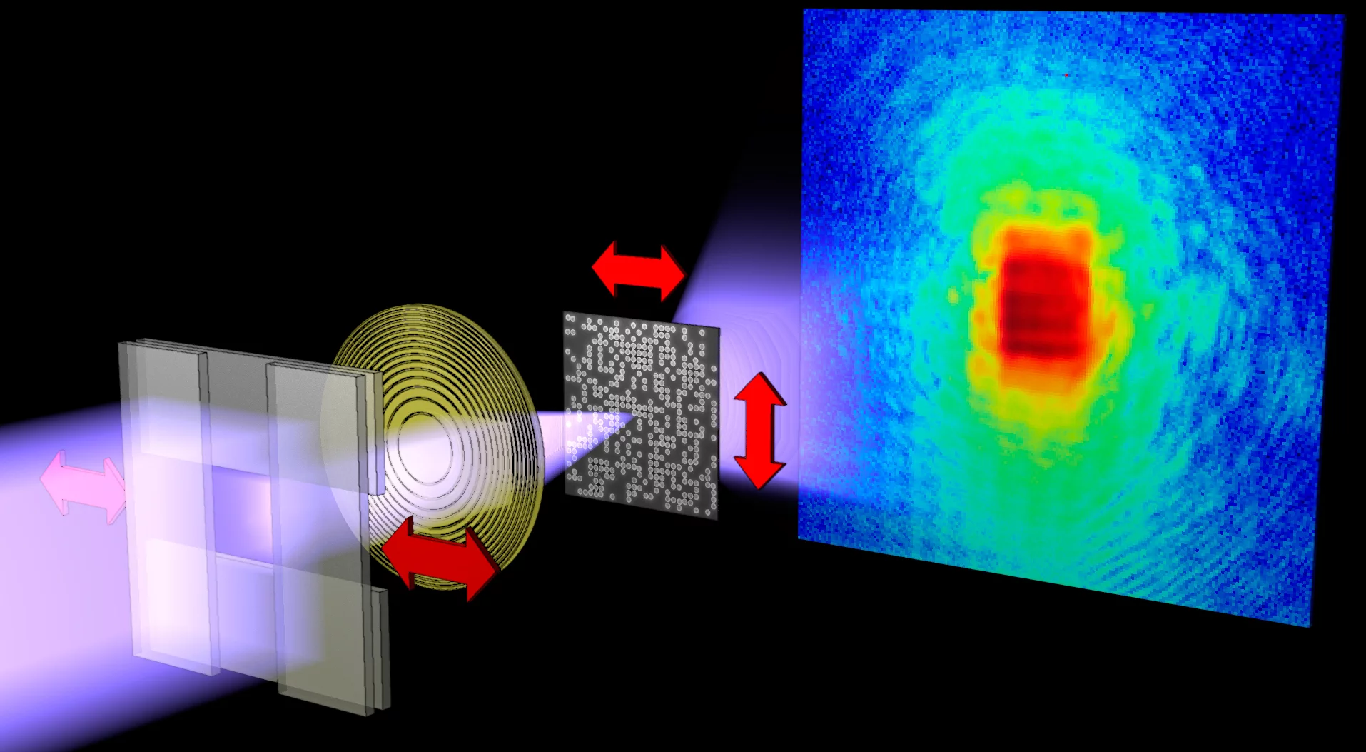
Microscopy using X-rays offers unique three-dimensional insights for the life and material sciences, e.g., into the architecture of biological cells, the porosity of concrete, or the domains of magnetic storage materials. Yet, investigating structures at the nanoscale, the requirements on the imaging apparatus bring enormous complications. Additionally, filters have to be applied in order to use only X rays with exactly the right properties, a particular wavelength for instance. “More importantly,” adds Andreas Menzel, scientist at the Paul Scherrer Institut, “unless light we use for the measurements is extremely well characterized, images can fail to render faithfully what we are looking at.”
Disentangling different wavelength contributions
Menzel and Pierre Thibault from the Technische Universität München developed an analysis method that allows objects to be imaged with high accuracy despite fluctuations or vibrations. Their method is based on a technique called “ptychography,” which was invented in the 1960s for microscopy using electrons and has further been developed during the last years to be a reliable high-resolution microscopy technique applicable also with X rays and visible light. The new results make now possible to disentangle accurately contributions of light with slightly different properties, such as different wavelengths. “Next to the immediate effects on imaging applications,” explains Pierre Thibault, “our analysis reveals deep connections to other well established fields of physics. Disciplines that have been seen as rather independent of imaging, such as quantum information processing, can benefit and cross-pollinate.”
Imaging fluctuations and mixtures
Perhaps most intriguingly, the newly developed technique can be used to image an entire new class of objects. “In addition to the imaging apparatus, we can allow the sample itself to fluctuate”, says Andreas Menzel. “Fluctuations that are too fast to be resolved by individual snap shots can thus be investigated.” One idea is to investigate magnetic materials with the magnetization of some domains fluctuating.
Confirmation by computer simulations
“We needed to convince ourselves that the images we produced did indeed represent the sample accurately and that the characterization of its dynamics was trustworthy,” elaborates Pierre Thibault. “To this task we used computer simulations that demonstrated that the method works for both instrumental and sample effects, such as flows, switching events, or quantum mixtures.” The analytical results presented in the study combine dynamics characterization with high-resolution imaging and are expected to facilitate fundamental studies, for instance, investigating the cross talk of individual bits on hard disks or the thermal fluctuations that limit the life time of such data storage.
About PSI
The Paul Scherrer Institute develops, builds and operates large, complex research facilities, and makes them available to the national and international research community. The Institute's own key research priorities are in the investigation of matter and material, energy and the environment; and human health. PSI is Switzerland's largest research institution, with 1500 members of staff and an annual budget of approximately 300 million CHF.
About Technische Universität München (TUM)
Technische Universität München (TUM) is one of Europe’s leading universities. It has roughly 480 professors, 9,000 academic and non-academic staff, and 32,000 students. It focuses on the engineering sciences, natural sciences, life sciences, medicine, and economic sciences. After winning numerous awards, it was selected as an “Excellence University” in 2006 and 2012 by the Science Council (Wissenschaftsrat) and the German Research Foundation (DFG). In both international and national rankings, TUM is rated as one of Germany’s top universities. TUM is dedicated to the ideal of a top-level research-oriented entrepreneurial university. The university's global presence includes offices in Beijing (China), Brussels (Belgium), Cairo (Egypt) and Sao Paulo (Brazil). The German Institute of Science and Technology (GIST), founded in 2002 in Singapore, is the first research campus of a German university abroad. www.tum.de
Contact
Dr. Andreas Menzel; Labor für Makromoleküle und Bioimaging;Paul Scherrer Institut, 5232 Villigen PSI, Switzerland
Phone: +41 56 310 3711; E-mail: andreas.menzel@psi.ch [German, English]
Dr. Pierre Thibault; Physikdepartment;
Technische Universität München, 85748 Garching, Germany
Phone: +49 (0)89 289 14397; E-mail: pierre.thibault@tum.de [French, English]
Original Publication
Reconstructing state mixtures from diffraction measurementsPierre Thibault & Andreas Menzel
Nature, 7. February 2013, DOI: 10.1038/nature11806