Associate Professor
Institute of Molecular Biology and Biophysics, ETH Zurich
Research Interests
Interactions between the cell and its environment, as well as between different cellular compartments, occur at the biological membranes. Extracellular signals are sensed by the receptors at the cell surface. These signals are then transmited across the membrane via multiple biomolecular interactions involving membrane-bound and soluble proteins, resulting in complex biochemical responses inside the cell. The process of signal transduction is critically important for physiology in health and disease. The research in my group is focused on understanding the molecular mechanisms of signal transduction. Using a multidisciplinary approach including methods of membrane protein biochemistry, biophysics and structural biology (including X-ray crystallography and cryo-EM), we aim to understand the structure-function relationships of membrane proteins and protein complexes involved in various aspects of cellular signalling. Among other topics, we are particularly interested in membrane protein complexes in two areas of cellular signalling: (i) the cyclic adenosine monophosphate (cAMP) pathway, (ii) gap junction intercellular communication.
Research Highlights
1. Structure and function of adenylyl cyclases: central enzymes in cAMP signaling
Membrane adenylyl cyclases (ACs) catalyze the conversion of ATP to the ubiquitous second messenger cAMP. As the major effector proteins of G protein-coupled receptors (GPCRs), ACs amplify diverse signals from outside of the cell and translate them into biochemical response inside the cell. Moreover, ACs are capable of integrating different signaling pathways, such as cAMP signaling and calcium signaling pathway. Our research has focused on detailed biochemical and structural studies of various membrane ACs, providing critical insights into their function. These include: (i) AC9 in its autoinhibited state bound to the stimulatory Gαs protein subunit (Qi et al., 2019), (ii) the mycobacterial membrane AC homologue Rv1625c/Cya (Mehta, Khanppnavar et al., 2022), and (iii) calcium/calmodulin-sensitive AC8 in complex with Gαs and calmodulin (Khanppnavar, Schuster et al., 2023). These studies establish a structural framework for understanding how membrane ACs and their macromolecular complexes with various partners initiate and regulate cAMP signaling.
Qi et al., Science, 2019: https://science.sciencemag.org/content/364/6438/389
Mehta, Khappnavar et al., eLife, 2022: https://elifesciences.org/articles/77032
Khanppnavar, Schuster et al., EMBO Reports, 2023: https://www.embopress.org/doi/full/10.1038/s44319-024-00076-y
2. Structural studies of connexin gap junction channels and hemichannels
Gap junction channels (GJCs), formed by connexins, facilitate intercellular communication by linking connexin hemichannels (HCs) on adjacent cells. These channels enable direct metabolic and electrical coupling between cells across various tissues, playing essential roles in numerous physiological and pathological processes. Using cryo-EM analysis combined with biochemistry, cell biology, and electrophysiology approaches, we investigate the molecular mechanisms underlying connexin channel function. Key findings include: (i) the structure of Cx43 in a closed state (Qi, Acosta Gutierrez et al., 2023), (ii) structural characterization of wild-type and Charcot-Marie-Tooth disease-linked mutants of Cx32 GJCs and HCs (Qi, Lavriha, Bayraktar et al., 2023), and (iii) identification of a novel mode of small molecule-mediated inhibition of Cx36 GJCs (Ding, Aureli et al., 2024). These studies provide valuable insights into connexin channel regulation and their roles in health and disease.
Qi, Acosta Gutierrez et al., eLife, 2023: https://elifesciences.org/articles/87616
Qi, Lavriha, Bayraktar et al., Sci Adv, 2023: https://www.science.org/doi/10.1126/sciadv.adh4890
Ding, Aureli et al., Cell Discov, 2024: https://www.nature.com/articles/s41421-024-00691-y
Key Publications
1. Ding X, Aureli S, Vaithia A, Lavriha P, Schuster D, Khanppnavar B, Li X, Blum TB, Picotti P, Gervasio FL, Korkhov VM. Structural basis of connexin-36 gap junction channel inhibition. Cell Discov. 2024 Jun 18;10(1):68. doi: 10.1038/s41421-024-00691-y.
2. Khanppnavar B, Schuster D, Lavriha P, Uliana F, Özel M, Mehta V, Leitner A, Picotti P, Korkhov VM. Regulatory sites of CaM-sensitive adenylyl cyclase AC8 revealed by cryo-EM and structural proteomics. EMBO Rep. 2024 Mar;25(3):1513-1540. doi: 10.1038/s44319-024-00076-y.
3. Qi C, Lavriha P, Bayraktar E, Vaithia A, Schuster D, Pannella M, Sala V, Picotti P, Bortolozzi M, Korkhov VM. Structures of wild-type and selected CMT1X mutant connexin 32 gap junction channels and hemichannels. Sci Adv. 2023 Sep;9(35):eadh4890. doi: 10.1126/sciadv.adh4890.
4. Qi C, Lavriha P, Mehta V, Khanppnavar B, Mohammed I, Li Y, Lazaratos M, Schaefer JV, Dreier B, Plückthun A, Bondar AN, Dessauer CW, Korkhov VM. Structural basis of adenylyl cyclase 9 activation. Nat Commun. 2022 Feb 24;13(1):1045. doi: 10.1038/s41467-022-28685-y.
5. Cannac F, Qi C, Falschlunger J, Hausmann G, Basler K, Korkhov VM. Cryo-EM structure of the Hedgehog release protein Dispatched. Sci Adv. 2020 Apr 15;6(16):eaay7928. doi: 10.1126/sciadv.aay7928.
6. Qi C, Di Minin G, Vercellino I, Wutz A, Korkhov VM. Structural basis of sterol recognition by human hedgehog receptor PTCH1. Sci Adv. 2019 Sep 18;5(9):eaaw6490. doi: 10.1126/sciadv.aaw6490.
7. Qi C, Sorrentino S, Medalia O, Korkhov VM. The structure of a membrane adenylyl cyclase bound to an activated stimulatory G protein. Science. 2019 Apr 26;364(6438):389-394. doi: 10.1126/science.aav0778.
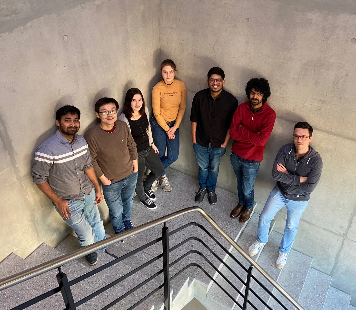
Group Members
Former Members
Weinert, Adriana (Postdoc)
Vercellino, Irene (Ph.D. Student)
Graeber, Elisabeth (Ph.D. Student)
Cannac, Fabien (Ph.D Student)
Oezel, Merve (Master/Ph.D Student)
Mehta, Ved (Ph.D Student)
Lavriha, Pia (Ph.D Student)
Schuster, Dina (Ph.D Student)
Publications
-
Khanppnavar B, Leka O, Pal SK, Korkhov VM, Kammerer RA
Cryo-EM structure of the botulinum neurotoxin A/SV2B complex and its implications for translocation
Nature Communications. 2025; 16(1): 1224 (16 pp.). https://doi.org/10.1038/s41467-025-56304-z
DORA PSI -
Ding X, Aureli S, Vaithia A, Lavriha P, Schuster D, Khanppnavar B, et al.
Structural basis of connexin-36 gap junction channel inhibition
Cell Discovery. 2024; 10(1): 68 (4 pp.). https://doi.org/10.1038/s41421-024-00691-y
DORA PSI -
Holfeld A, Schuster D, Sesterhenn F, Gillingham AK, Stalder P, Haenseler W, et al.
Systematic identification of structure-specific protein–protein interactions
Molecular Systems Biology. 2024; 20: 651-675. https://doi.org/10.1038/s44320-024-00037-6
DORA PSI -
Khanppnavar B, Schuster D, Lavriha P, Uliana F, Özel M, Mehta V, et al.
Regulatory sites of CaM-sensitive adenylyl cyclase AC8 revealed by cryo-EM and structural proteomics
EMBO Reports. 2024; 25: 1513-1540. https://doi.org/10.1038/s44319-024-00076-y
DORA PSI -
Khanppnavar B, Choo JPS, Hagedoorn PL, Smolentsev G, Štefanić S, Kumaran S, et al.
Structural basis of the Meinwald rearrangement catalysed by styrene oxide isomerase
Nature Chemistry. 2024; 16: 1496-1504. https://doi.org/10.1038/s41557-024-01523-y
DORA PSI -
Lavriha P, Qi C, Korkhov VM
Expression, purification, and nanodisc reconstitution of connexin-43 hemichannels for structural characterization by cryo-electron microscopyp
In: Mammano F, Retamal M, eds. Connexin hemichannels. Methods and protocols. Methods in molecular biology. New York, NY: Springer Nature; 2024:29-43. https://doi.org/10.1007/978-1-0716-3842-2_3
DORA PSI -
Lavriha P, Han Y, Ding X, Schuster D, Qi C, Vaithia A, et al.
Mechanism of connexin channel inhibition by mefloquine and 2-aminoethoxydiphenyl borate
PLoS One. 2024; 19(12): e0315510 (15 pp.). https://doi.org/10.1371/journal.pone.0315510
DORA PSI -
Saha S, Khanppnavar B, Maharana J, Kim H, Carino CMC, Daly C, et al.
Molecular mechanism of distinct chemokine engagement and functional divergence of the human Duffy antigen receptor
Cell. 2024; 187(17): 4751-4769.e25. https://doi.org/10.1016/j.cell.2024.07.005
DORA PSI -
Schuster D, Khanppnavar B, Kantarci I, Mehta V, Korkhov VM
Structural insights into membrane adenylyl cyclases, initiators of cAMP signaling
Trends in Biochemical Sciences. 2024; 49(2): 156-168. https://doi.org/10.1016/j.tibs.2023.12.002
DORA PSI -
Barret DCA, Schuster D, Rodrigues MJ, Leitner A, Picotti P, Schertler GFX, et al.
Structural basis of calmodulin modulation of the rod cyclic nucleotide-gated channel
Proceedings of the National Academy of Sciences of the United States of America PNAS. 2023; 120(15): e2300309120 (10 pp.). https://doi.org/10.1073/pnas.2300309120
DORA PSI -
Qi C, Gutierrez SA, Lavriha P, Othman A, Lopez-Pigozzi D, Bayraktar E, et al.
Structure of the connexin-43 gap junction channel in a putative closed state
eLife. 2023; 12: RP87616 (27 pp.). https://doi.org/10.7554/eLife.87616
DORA PSI -
Qi C, Lavriha P, Bayraktar E, Vaithia A, Schuster D, Pannella M, et al.
Structures of wild-type and selected CMT1X mutant connexin 32 gap junction channels and hemichannels
Science Advances. 2023; 9(35): eadh4890 (14 pp.). https://doi.org/10.1126/sciadv.adh4890
DORA PSI -
Khanppnavar B, Maier J, Herborg F, Gradisch R, Lazzarin E, Luethi D, et al.
Structural basis of organic cation transporter-3 inhibition
Nature Communications. 2022; 13: 6714 (13 pp.). https://doi.org/10.1038/s41467-022-34284-8
DORA PSI -
Mehta V, Khanppnavar B, Schuster D, Kantarci I, Vercellino I, Kosturanova A, et al.
Structure of Mycobacterium tuberculosis Cya, an evolutionary ancestor of the mammalian membrane adenylyl cyclases
eLife. 2022; 11: e77032 (21 pp.). https://doi.org/10.7554/ELIFE.77032
DORA PSI -
Qi C, Lavriha P, Mehta V, Khanppnavar B, Mohammed I, Li Y, et al.
Structural basis of adenylyl cyclase 9 activation
Nature Communications. 2022; 13(1): 1045 (11 pp.). https://doi.org/10.1038/s41467-022-28685-y
DORA PSI -
Lentini G, Ben Chaabene R, Vadas O, Ramakrishnan C, Mukherjee B, Mehta V, et al.
Structural insights into an atypical secretory pathway kinase crucial for Toxoplasma gondii invasion
Nature Communications. 2021; 12(1): 3788 (17 pp.). https://doi.org/10.1038/s41467-021-24083-y
DORA PSI -
Zhang X, Pizzoni A, Hong K, Naim N, Qi C, Korkhov V, et al.
CAP1 binds and activates adenylyl cyclase in mammalian cells
Proceedings of the National Academy of Sciences of the United States of America PNAS. 2021; 118(24): e2024576118 (10 pp.). https://doi.org/10.1073/pnas.2024576118
DORA PSI -
Cannac F, Qi C, Falschlunger J, Hausmann G, Basler K, Korkhov VM
Cryo-EM structure of the Hedgehog release protein Dispatched
Science Advances. 2020; 6(16): eaay7928 (8 pp.). https://doi.org/10.1126/sciadv.aay7928
DORA PSI -
Graeber E, Korkhov VM
Affinity purification of membrane proteins
In: Perez C, Maier T, eds. Expression, purification, and structural biology of membrane proteins. Methods in molecular biology. New York: Humana; 2020:129-137. https://doi.org/10.1007/978-1-0716-0373-4_9
DORA PSI -
Khannpnavar B, Mehta V, Qi C, Korkhov V
Structure and function of adenylyl cyclases, key enzymes in cellular signaling
Current Opinion in Structural Biology. 2020; 63: 34-41. https://doi.org/10.1016/j.sbi.2020.03.003
DORA PSI -
Graeber E, Korkhov VM
Characterisation of the ligand binding sites in the translocator protein TSPO using the chimeric bacterial-mammalian constructs
Protein Expression and Purification. 2019; 164: 105456 (10 pp.). https://doi.org/10.1016/j.pep.2019.105456
DORA PSI -
Qi C, Minin GD, Vercellino I, Wutz A, Korkhov VM
Structural basis of sterol recognition by human hedgehog receptor PTCH1
Science Advances. 2019; 5(9): eaaw6490 (9 pp.). https://doi.org/10.1126/sciadv.aaw6490
DORA PSI -
Qi C, Sorrentino S, Medalia O, Korkhov VM
The structure of a membrane adenylyl cyclase bound to an activated stimulatory G protein
Science. 2019; 364(6438): 389-394. https://doi.org/10.1126/science.aav0778
DORA PSI -
Graeber E, Korkhov VM
Expression and purification of the mammalian translocator protein for structural studies
PLoS One. 2018; 13(6): e0198832 (17 pp.). https://doi.org/10.1371/journal.pone.0198832
DORA PSI -
Mireku SA, Ruetz M, Zhou T, Korkhov VM, Kräutler B, Locher KP
Conformational change of a tryptophan residue in BtuF facilitates binding and transport of cobinamide by the vitamin B12 transporter BtuCD-F
Scientific Reports. 2017; 7: 41575 (11 pp.). https://doi.org/10.1038/srep41575
DORA PSI -
Vercellino I, Rezabkova L, Olieric V, Polyhach Y, Weinert T, Kammerer RA, et al.
Role of the nucleotidyl cyclase helical domain in catalytically active dimer formation
Proceedings of the National Academy of Sciences of the United States of America PNAS. 2017; 114(46): E9821-E9828. https://doi.org/10.1073/pnas.1712621114
DORA PSI -
Korkhov VM, Mireku SA, Veprintsev DB, Locher KP
Structure of AMP-PNP-bound BtuCD and mechanism of ATP-powered vitamin B12 transport by BtuCD-F
Nature Structural and Molecular Biology. 2014; 21(12): 1097-1099. https://doi.org/10.1038/nsmb.2918
DORA PSI -
Chen F, Gerber S, Heuser K, Korkhov VM, Lizak C, Mireku S, et al.
High-mass matrix-assisted laser desorption ionization-mass spectrometry of integral membrane proteins and their complexes
Analytical Chemistry. 2013; 85(7): 3483-3488. https://doi.org/10.1021/ac4000943
DORA PSI -
Korkhov VM, Mireku SA, Hvorup RN, Locher KP
Asymmetric states of vitamin B12 transporter BtuCD are not discriminated by its cognate substrate binding protein BtuF
FEBS Letters. 2012; 586(7): 972-976. https://doi.org/10.1016/j.febslet.2012.02.042
DORA PSI -
Korkhov VM, Mireku SA, Locher KP
Structure of AMP-PNP-bound vitamin B12 transporter BtuCD-F
Nature. 2012; 490(7420): 367-372. https://doi.org/10.1038/nature11442
DORA PSI
