Current Research Topics
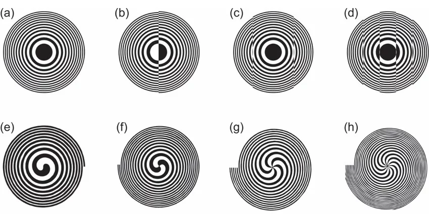
X-ray diffractive optics offer great versatility to manipulate X-ray beams for interesting scientific applications. Tailored X-ray diffractive optics, such as the different type of Fresnel zone plates shown in figure 1, can perform advanced optical functions and manipulate the wavefront and focus of the X-ray beams.
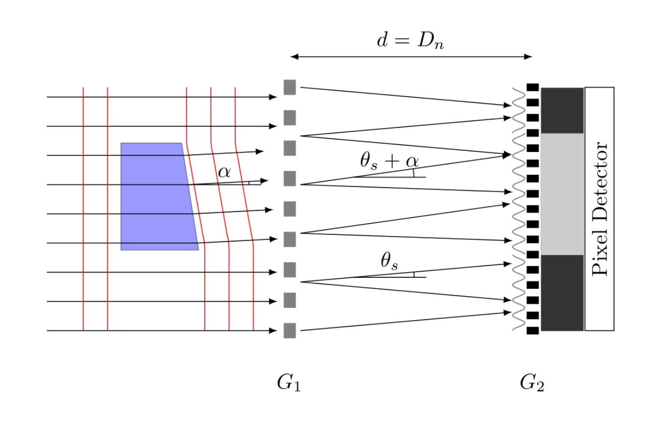
On the other hand, grating-based X-ray interferometry (see figure 2) has ventured into many areas of X-ray imaging and wavefront metrology, since its first demonstration by LMN researchers in 2002 [1]. A typical grating interferometer consists of a phase shifting grating G1 made made of silicon which diffracts the incident radiation mainly into a first and minus first diffraction order that propagate at angles +θ and -θ with respect to their original direction of propagation. These two orders interfere at a distance d downstream, forming a periodic interference pattern that can be analyzed by an absorbing grating G2made of gold and a camera. Any refracting object in the beam, such as the wedge shown in figure 2, a biomedical or material science sample in imaging [2] or an X-ray optical element in metrology, leads to a locally modified propagation direction of the X-ray beam. This beam angle α leads to a local lateral shift of the interference pattern that can be detected by the combination of analyzer grating and camera. From this shift, the refraction angle α can be quantitatively determined.
Publications
- C. David, B. Nöhammer, H.H. Solak, E. Ziegler, Differential x-ray phase contrast imaging using a shearing interferometer, Applied Physics Letters 81, no. 17 (2002) p. 3287-3290.
- T. Weitkamp, B. Nöhammer, A. Diaz, C. David, E. Ziegler, X-ray wavefront analysis and optics characterization with a grating interferometer, Applied Physics Letters 86 (2005) p. 054101-054103.


