X-ray Nano-Optics

XRO start.jpg

The X-ray Nano-Optics group of LXN works on various fields of research involving the control of x-rays with ultimate precision. We develop instrumentation for large scale facilities such as synchrotrons and x-ray free electron lasers (XFELs), by applying nanolithography techniques. This includes x-ray diffractive optics such as Fresnel zone plate lenses for imaging and probing of matter on a micro- and nanometer scale. Our optics are used at many synchrotron beam lines worldwide and hold the resolution record in x-ray microscopy. For applications that do not require ultimate resolution, we pursue novel approaches to obtain very high diffraction efficiencies.

Interferometric imaging techniques using hard x-rays also rely on specialized micro-fabricated gratings. The possibility to use this technique not only with synchrotron radiation but also with incoherent x-rays from tube sources makes the technique interesting for commercial applications. The extreme sensitivity of grating interferometry also provides a powerful tool for x-ray optics metrology and wavefront sensing.

The development of a new generation of x-ray sources based on the x-ray free-electron laser (XFEL) principle have triggered the development of specialized diffractive optics. In order to withstand the extreme power levels of XFELs, we make Fresnel zone plates based on diamond substrates. Similar devices are made for applications such as spectral monitoring or beam splitting. The latter is used to build multiple split-and-delay lines for ultra-fast pump-probe experiments with unprecedented timing precision.

For the fabrication of these devices, the X-Ray Optics and Applications Group runs LMN’s high performance electron-beam lithography tool Vistec EBPG 5000PlusES, that is also used by many other internal and external research groups.


Publications

For full list of Publications of the X-ray Optics and Applications group (since 2005) see: DORA


Highlights

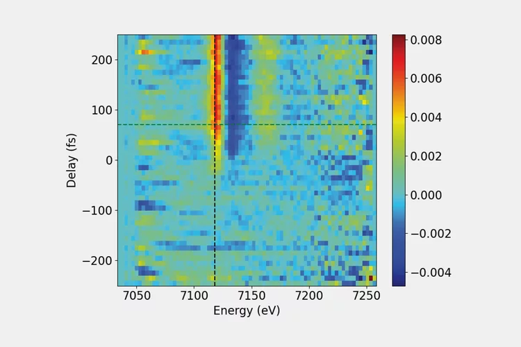
Delay vs. photon energy map of an ammonium iron(III) oxalate solution. The large bandwidth emission covers the full XANES range, making time-consuming monochromator scans obsolete.

Efficient transient X-ray absorption spectroscopy

By combining the unique large bandwidth emission mode of SwissFEL’s ARAMIS undulator and diffractive X-ray optics made of diamond, we have demonstrated a new method for time-resolved X-ray absorption near edge structure (XANES) spectroscopy that enables faster data acquisition and requires smaller sample quantities for high-quality data. 

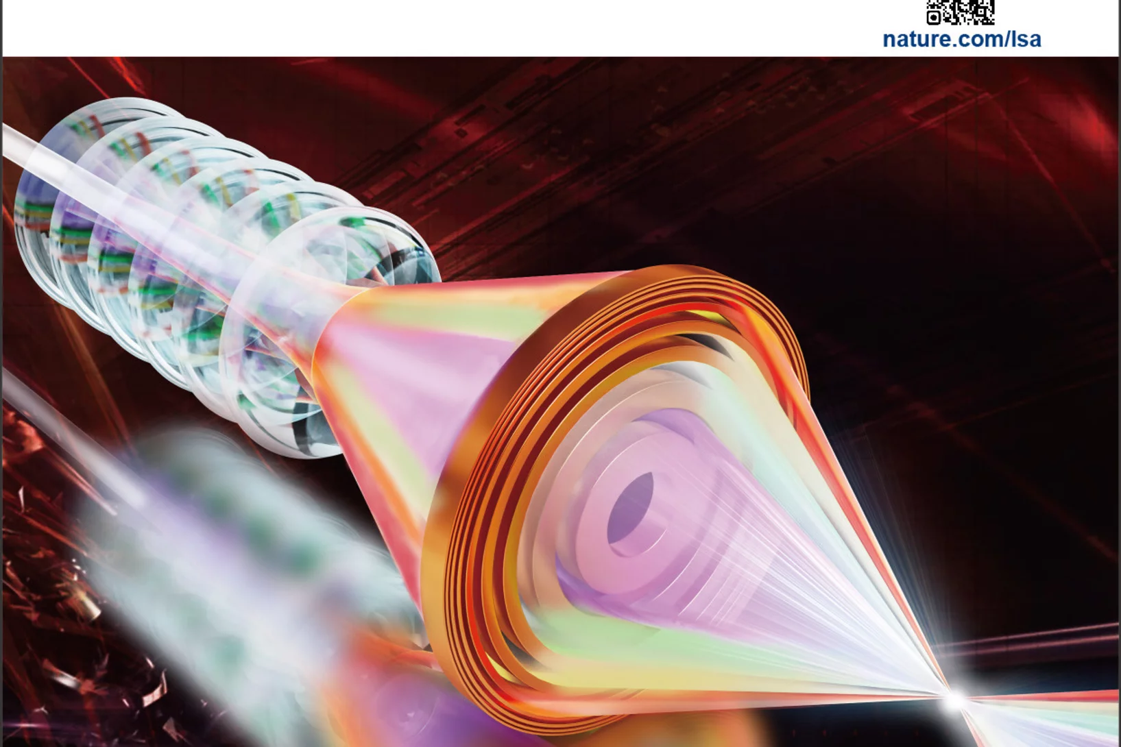
Apochromatic X-ray focusing Editor Highlight

Apochromatic X-ray focusing as Editor's Highlight in Light: Science & Applications

Our recent work on the 1st demonstration of apochromatic X-ray lenses has been selected as an Editor's Highlight in Light: Science & Applications.

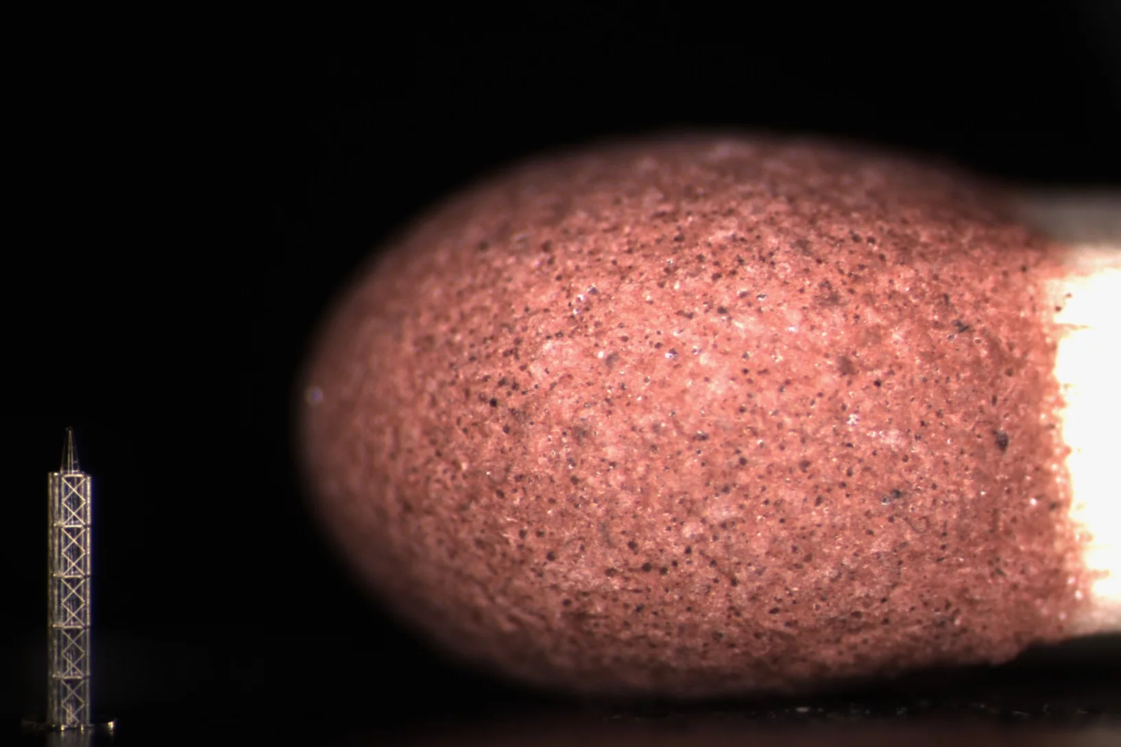
Photograph of the refractive X-ray lens

Apochromatic X-ray focusing

A team of scientists from the Paul Scherrer Institut, the University of Basel and DESY have demonstrated the first-ever realization of apochromatic X-ray focusing using a tailored combination of a refractive lens and a Fresnel zone plate. This innovative approach enables the correction of the chromatic aberration suffered by both refractive and diffractive lenses over a wide range of X-ray energies. This groundbreaking development in X-ray optics have been just published in the scientific journal Light: Science & Applications.

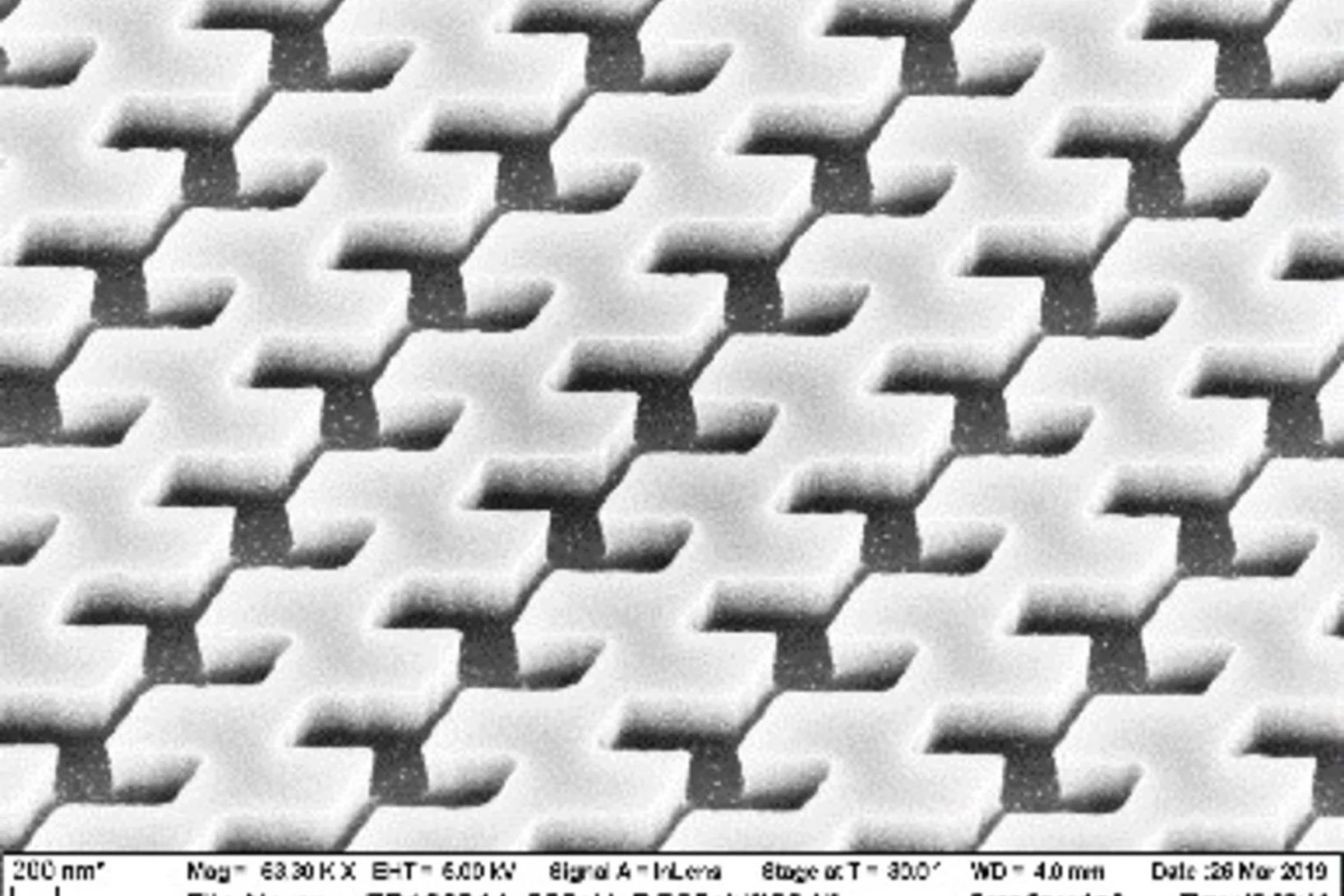
diamond_grantings_teaser

Diamond gratings for XFEL amplitude-splitting delay line

A split-and-delay line for XFEL pulses has been built and successfully tested by a team of researchers at the Linac Coherent Light Source. Key X-ray optical elements are two diamond diffraction gratings made at the Paul Scherrer Institut that are used to split and later recombine the intense ultrashort X-ray laser pulses for time-resolved measurements.

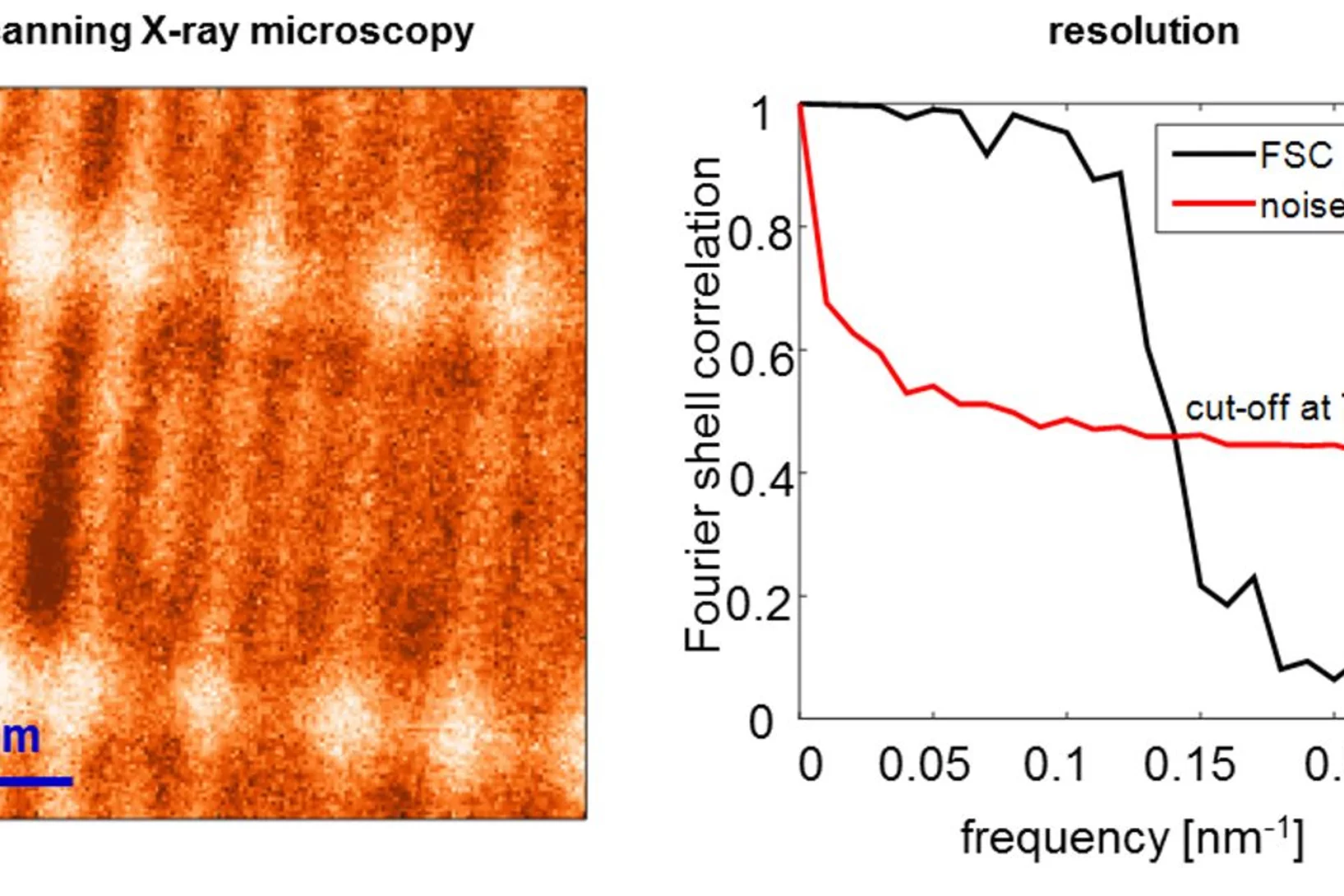
High-resolution X-ray microscopy of a test pattern with 9 nm line width

World Record: 7 nm Resolution in Scanning Soft X-ray Microscopy

During the past decade, scientists have put high effort to achieve sub-10 nm resolution in X-ray microscopy. Recent developments in high-resolution lithography-based diffractive optics, combined with the extreme stability and precision of the PolLux and HERMES scanning X-ray microscopes, resulted now in a so far unreached resolution of seven nanometers in scanning soft X-ray microscopy. Utilizing this highly precise microscopy technique with the X-ray magnetic circular dichroism effect, dimensionality effects in an ensemble of interacting magnetic nanoparticles can be revealed.

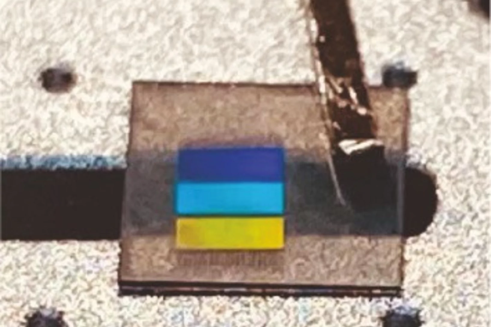
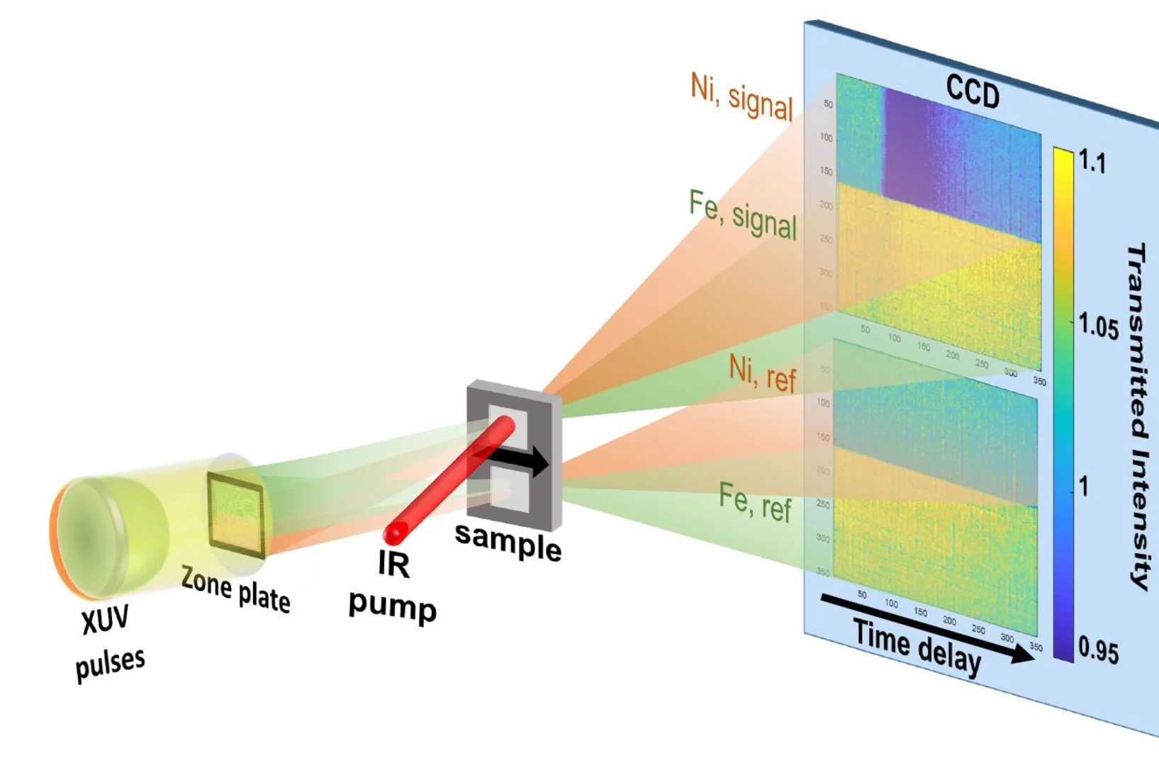
Two-color, twin-focus zone plate

Two-color snapshots of ultrafast charge and spin dynamics

In a joint research effort, an international team of scientists lead by Emmanuelle Jal (Sorbonne Université) performed a time-resolved experiment at the FERMI free-electron laser to disclose the dynamic behavior of two magnetic element of a compount material in only one snapshot. The X-ray Optics and Applications group developed a dedicated optical element for this experiment that is usable with two different photon energies (colors) simultaneously.

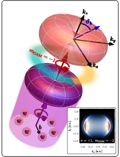
OAM Imprinting on He Atoms

Photoelectric Effect with a Twist

In a joint research effort, an international team of scientists lead by Prof. Giovanni de Ninno (University of Nova Gorica, Elettra Sincrotrone Trieste) now demonstrated that an OAM-dependent dichroic effect can be observed on photoelectrons. The photoelectrons are released from a sample of He atoms that is excited by the strong extreme ultraviolet light pulses from the FERMI free electron laser, whereas the orbtial momentum is imprinted with an intense infrared laser pulse. The X-ray Optics and Applications group of PSI supported the team with their experience in the creation of OAM beams and during the experiments.

Novel Optics

Novel optics enable better X-ray Free Electron Laser experiments

Our research on multifocus off-axis zone plates was accepted in “Optica”, the highest impact journal of the Optical Society of America. In the paper we report on different ways to combine focusing and beam-splitting functionalities in one single optical element.

logo of xraynanotech

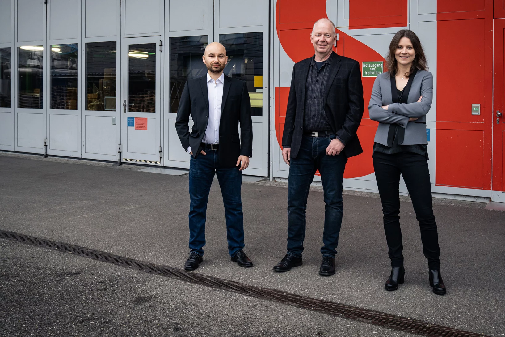
Florian Döring received the PSI Founder Fellowship

A new PSI spin-off is on the horizon: Dr. Florian Döring, PostDoc in the Laboratory for Micro- and Nanotechnology, received a PSI Founder Fellowship at Park Innovaare.

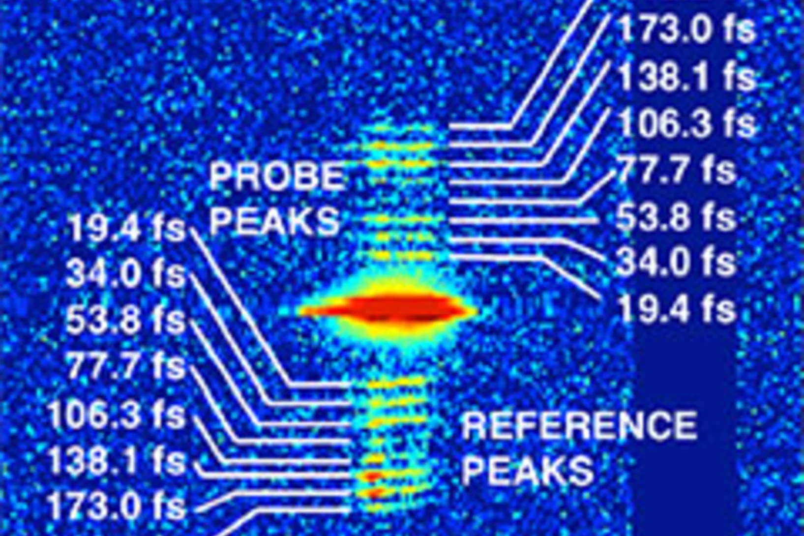
Demonstration of femtosecond X-ray pump X-ray probe diffraction on protein crystals

Our experiments, published in the September issue of Structural Dynamics, demonstrate the feasibility of time-resolved pump-multiprobe X-ray diffraction experiments on protein crystals using a split-and-delay setup which was temporarily installed at the LCLS X-ray Free Electron Laser.